Contents
The Light Microscope
Hooke’s Cell


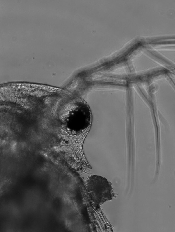
The father of Microbiology: van Leeuwenhoek

Though van Leeuwenhoek’s apparatus was simple, the magnifying power of his lenses and his curiosity enabled him to perform great scientific observations on the the microscopic world. He was ridiculed for fabricating his observations of protists at first. Ever the scientist, van Leeuwenhoek examined samples of his own diarrhea to discover Giardia intestinalis. While he did not make the connection of the causative nature of this microorganism, he described the details of the way this organism could propel itself through the medium in great detail.
.jpg)
Modern Compound Microscope
Unlike van Leeuwenhoek’s single lens microscope, we now combine the magnifying power of multiple lenses in what is called a compound microscope.

- Ocular lens or eyepiece
- Nose Piece/ Lens Carousel
- Objective lens
- Course Focus Knob
- Fine Focus Knob
- Stage
- lamp
- condenser
- stage control
Using the Light Microscope
Microscopic World

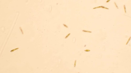
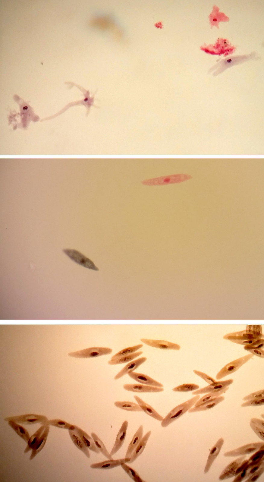
Scale

- Discover more about scale and microscopy at this link http://learn.genetics.utah.edu/content/cells/scale/
Tags: guided inquiry, quantitative reasoning, visual communication

