For the most part, mammals have gender determined by the presence of the Y chromosome. This chromosome is gene poor and a specific area called sex determining region on Y (SRY) is responsible for the initiation of the male sex determination. The X-chromosome is rich in genes while the Y-chromosome is a gene desert. The presence of an X-chromosome is absolutely necessary to produce a viable life form and the default gender of mammals is traditionally female.
The human X-chromosomeThe minuscule human Y-chromosome
Chromosomal painting techniques can reveal the gender origin of mammalian cells. By using fluorescent marker sequences that can hybridize specifically to X or Y chromosomes through Fluorescence In Situ Hybridization (FISH), gender can be identified in cells.
Recombinant DNA technology has many uses in basic scientific research to better understand the nature of of living things. As a tool, recombinant DNA technology can be used to express proteins towards medical applications. Prior to biotechnology, type I diabetes (insulin-dependent) was treated by injection of insulin isolated from the pancreas of pigs. With the ability to express human proteins inside bacteria, yeast and other cells, sacrificing pigs for porcine insulin is no longer necessary.
Bacterial expression vector pGEX-3X contains the AmpR gene, origin of replication, MCS downstream of the hybrid lac/trp promoter (tac) and the coding sequence for glutathione-S-transferase (GST). GST acts as a tag that is fused directly with the protein from the gene of interest and used to purify the protein with a glutathione resin.
Bacteria or other cells can be engineered to express proteins through the process of cloning and transformation. Bacteria are advantageous because of their rapid life cycle and ease of growth. A bacterial expression vector contains the basic plasmid features: origin of replication as well as antibiotic resistance gene. Often, an affinity tag will be used to aid in purification of the protein. An example in the vector above shows the GST (glutathione-s-transferase) tag that can be purified with glutathione resin. Expression is only the first problem since bacteria are also synthesizing proteins that are required for the bacteria to grow and divide. Injecting these proteins in addition to insulin would cause an immune reaction that could be deadly. Therefore, it is required that overexpressed proteins be purified and isolated from other undesirable proteins.
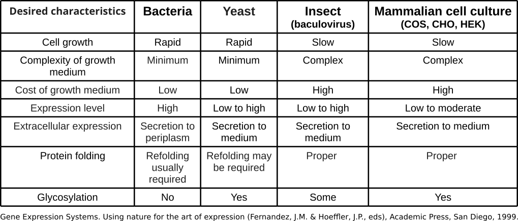
Criteria for Choosing an Expression System
Protein expression systems have inherent advantages and disadvantages. The table above summarizes the comparison of the various cellular systems of production (Fernandez & Hoeffler, 1999).
A bioreactor or fermenter that is used to grow large amounts of bacteria for the production of protein.
Purification
Different methods of isolation can be applied depending on the properties of the protein. Ion exchange chromatography is useful if the protein of interest has a specific charge that will interact with a resin packed with the opposite charge.
Immunoprecipitation
Immunoprecitation: Column is packed with Protein-A agarose which binds to antibodies. Cell lysates are then loaded onto the columns where they flow through and are allowed to interact with the antibody. Washes are performed to remove the non-specifically bound proteins. An elution buffer is used to disrupt the interaction of the antibody to the protein target.
Affinity Purification
Affinity purification employs the use of specific antibodies that bind to the protein of interest very tightly to retain it on a column. With these techniques, the protein retained on the resin is washed numerous times to remove other proteins that are non-specifically sticking. A change in pH or ionic conditions then is used to disrupt the interaction with the resin and elute the proteins from the column. Proteins that are engineered to contain tags can be purified by antibodies specific to those tags. Also, the addition of 6 or more consecutive Histidine residues to the end of a protein make them susceptible to purification with Nickel-NTA resin or Cobalt purification. In these cases, the 6XHis tag associates with these metal ions on the resin and are selectively adhered.
Nickel NTA resin coordinating the capture of a 6His tagged protein
Size Exclusion
Most of you are familiar with water purification filters. Before using these filters, you soak them in water and dark residue leaks out. This dark residue is activated charcoal. The activated charcoal has tiny microscopic pores that trap small items like ions and other particles. The primary goal of these filters is to remove metals and chlorine that are found in tap water. The porous nature of activated charcoal renders it useful for trapping molecules in water purification systems . The process used to trap these small particles is called size exclusion. Unlike agarose gel electrophoresis where the smaller particles navigate through the matrix faster, size exclusion resins trap the smaller molecules.
The smaller the molecule, the longer they spend within the pores as they traverse through the matrix.
Significance of Purification
All injectible drugs must be clean of endotoxins from bacteria. Purification of the protein of interest from bacterial lysates removes the dangerous pathogenic materials from that would otherwise activate host immune reactivity.
The horseshoe crab (Limulus polyphemus) performs a special function in the ecosystem by providing eggs for migratory birds to feed on. This organism also houses a special cell type in its hemolymph. The limulus amoebocyte lysate (LAL) test is the most sensitive assay of detecting endotoxins from bacteria. Amoebocytes are collected from these organisms for use on testing batches of injectible drugs to ensure proper purification and safety.
Genetic modification of organisms has been occurring through human manipulation since the beginning of agriculture. Humans selectively bred crops and livestock to propagate desirable traits in a process termed artificial selection. The original grass that gave rise to domesticated corn called teosinte hardly resembles what we think of when imagining modern maize.
Teosinte, the progenitor of maize. Corn came about due to selective breeding.
Variation: Crop domestication
Selective breeding can yield a variety of features even within the same species. Below is selection of vegetables of the species Brassica oleracea that have been developed into different varieties over the course of agricultural history.
Cabbage: Brassica oleracea var. capitataBroccoli: Brassica oleracea var. italicaKohlrabi: Brassica oleracea var. gongylodesRomanesco: Brassica oleracea var. botrytis
Variation: Animal domestication
Companion animals like dogs underwent thousands of years of domestication and selection for traits that were desirable for different circumstances. A high degree of morphological diversity exists between dog breeds and their ancestral grey wolf progenitor.
Genetic Manipulation (engineered)
Artificial selection takes multiple generations over a long period of time. With the advent of recombinant DNA and biotechnology, scientists can now genetically modify organisms through introduction of foreign genes to provide desirable characteristics within one generation. This process does not require traits to naturally arise in a species.
GloFish are transgenic zebra fish (Danio rerio) expressing variants of GFP. Bottom features a wild-type fish.GloFish® are novelty pets that have the insertion of various cnidarian fluorescent protein genes into the genome. These fish were released in the United States in 2003 and have subsequently been developed in red, orange, and blue varieties. Black tetras and tiger barbs are also now available.
Black tetra (Gymnocorymbus ternetzi) GloFishWild-type Black Tetra
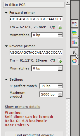
The PCR amplification of the mitochondrial control region
There are 2 hypervariable regions within the control region of the mitochondria. This exercise amplifies just one of these. For more definitive results, both should be amplified and sequenced. This exercise will permit us to have a rough idea of the origins of our maternal line and we will be able to attribute ourselves to various tribes throughout the world. The human mitochondrial genome (genbank file).
Forward Primer5’-TTAACTCCACCATTAGCACC-3’
Reverse Primer5’-GAGGATGGTGGTCAAGGGAC-3’
PCR the previously extract DNA samples
Pour 2% agarose into casting apparatus in refrigerator
2 gels per class need to be made → 100ml of TBE with 2g agarose
add 5μl SYBR safe solution into the molten agarose before casting
place 2 sets of combs into the gel → at one end and in the middle
load gel with DNA ladder and PCR
Run gel at 120V for 20 minutes
Visualize on UV transilluminator
Document with camera to verify amplification
The instructor will submit the viable reactions for sequencing
In addition to the 23 chromosomes inherited from mother and 23 chromosomes inherited from father, humans have an additional genome that is only inherited from the mother. This genome comes from the endosymbiotic organelle, the mitochondrion.
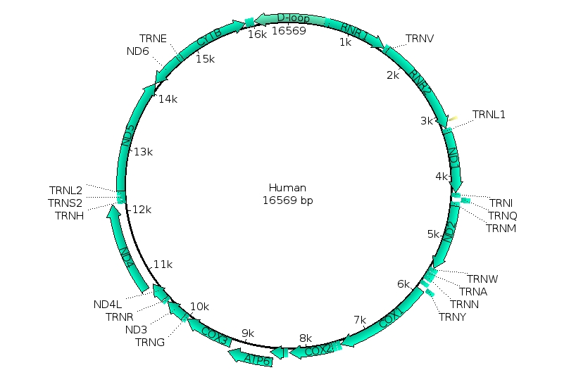
Mitochondria are thought to have arisen in the eukaryotic line when bacteria capable of detoxifying the deadly effects of atmospheric oxygen were engulfed by a eukaryote that did not proceed to consume it. Over the course of time, these formerly free-living bacteria became dependent on the eukaryotic cell environment while providing the benefit to the host cell of aerobic respiration. Hallmarks of this endosymbiotic event include: the inner prokaryotic membrane surrounded by the outer eukaryotic membrane, the presence of prokaryotic ribosomes and most significantly, the circular prokaryotic chromosome. Mitochondria still replicate independently of the host cell but can not survive outside of this cellular environment. Animal mitochondria have the simplest genomes of all mitochondrial genomes, ranging from 11-28kb. The human mitochondrial genome consists of 37 genes which are almost all devoted to processing ATP through oxidative phosphorylation.
Human mitochondrial genome
The human mitochondrial genome (genbank file) consists of 16,569 nucleotides (16.6kb). While most of this 16.6kb genome consists of protein encoding genes, approximately 1.2kb non-coding DNA takes part in signals that control the expression of these genes and replication processes. It is the area of DNA where the double-strandedness is displaced and having the name D-loop (displacement loop). Mutations in this area generally have very little effect on the functioning of the mitochondria. Because of this reduced selection pressure on this area, this control region is also referred to as the hypervariable region. This hypervariable region actually has 10 times more SNPs than the nuclear genome. Due to this abundance of mutations, it is possible to track down the maternal line of an individual. Why just maternal? The human oocyte contains many mitochondria while sperm cells only contain mitochondria that power the flagellar motion. Upon fertilization, the flagellum and the associated mitochondria are lost, leaving the zygote with only maternal mitochondria.
The cluster of SNPs found in the mitochondrial control region are linked and are always inherited together. Because of the lack of paternal contribution, this linkage is referred to as a haplotype, or “half-type”. Tracking these polymorphic haplotypes, a family tree of humans was developed in the 1980s which concluded that humans arose from a metaphorical “Mitochondrial Eve” 200,000 years ago. As a metaphor to the Biblical Eve, this alludes to an origin but unlike the Biblical event, this does not mean that it was a single woman that gave rise to all of modern humanity. On the contrary, the metaphor merely indicates that a series of females; sisters and cousins, of this line gave rise to modern humans.
Migration map of mitochondrial haplogroups. Numbers represent 1000 years ago.
The use of mitochondria for this analysis provides great flexibility, especially from ancient sources. Unlike the nuclear genome which only has 2 copies of DNA per cell, the mitochondria are abundant in number and provide many copies of genome per cell. Ancient sources of DNA in fossils will most often have degradation of the DNA. The mitochondrial genome is just as likely to undergo degradation over time, however the high copy number allows for gaps to be filled in easily. SNPs do not alter the overall size of the hypervariable region, therefore amplification by PCR can not resolve these differences based on agarose gel migration. However, amplicons (amplified copies) can be sent for sequencing whereby each nucleotide can be called out in succession and reveal the specific SNPs.
Alu’s are unique SINEs that appear in the primate lineage and reveal the lineage and diversification of primates. While retrotransposons can disrupt gene (as in some cases of hemophilia), they often land outside of genes or within introns without effect. One example of a non-disruptive Alu element in humans is found in the location called PV92 on chromosome 16. This element is of the youngest subfamily of Alu, called Ya5.
Since PV92 does not cause any deleterious effects, it can be used as a non-selected marker to illustrate lineage. Some people have an Alu element int his location while others do not. The presence or absence of this marker is viewed as an allele. This lab uses primer that flank the location of the Alu insertion that span 416 bp. If an Alu is present, the amplified DNA will be 300bp larger (the size of an Alu) at 731bp.
Drawings dating to approximately 30,000 years ago in the Chauvet CaveWhat constitutes being human? Many will point at a cultural identity and leaving long-standing remnants of that culture. Such prehistorical artifacts like cave drawings and tools provide an anthropological framework for identifying what it is to be human, but the biological identity remains locked in the history of our DNA.
Spear points of the Clovis Culture in the Americas dating to approximately 13,000 years ago.
The Great Apes
Phylogenetic tree generated with Cytochrome Oxidase I (COI) genes.
Homo sapiens represent a branch of primates in the line of Great Apes. The family of Great Apes consists of four extant genera: Homo, Pan, Gorilla, Pongo. Karyotype analysis (Yunis et al., 1982) reveals a shared genomic structure between the Great Apes. While humans have 46 chromosomes, the other Great Apes have 48. Molecular evidence at the DNA level indicates that Human Chromosome 2 is a fusion of 2 individual chromosomes. In the other Great Apes, these 2 Chromosomes are referred to as 2p and 2q to illustrate their synteny to the human counterpart.
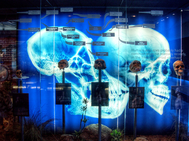
The Pan-Homo divergence. A display at the Cradle of Humankind illuminates the skulls of two extant Hominini with a series of model fossils from the Hominina subtribe of Austrolopithecina and Homo
Chimpanzees (Pan) are the closest living relatives to modern humans. It is commonly cited that less than 2% differences in their nucleotide sequences exist with humans (Chimpanzee Sequencing and Analysis Consortium, 2005). More recent findings in comparing the complement of genes (including duplication and gene loss events) now describes the difference in genomes at about 6% (Demuth JP, et al., 2006).
The Genus Homo
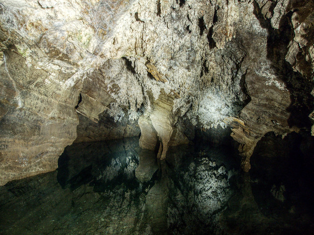
An underground lake at inside the Sterkfontein Cave system at the Cradle of Humankind (South Africa)
The rise of the human lineage is thought to arise in Africa. Fossils of Austroloptihs (southern apes) found in death traps, like those at the Cradle of Humankind, reveal a historical record of organisms inhabiting the landscape. The breaks in the ceiling of the caves provide opportunities for animals to fall inside these caves to their death. The limestone deposits of the caves serve as an environment for fossilization and mineralization of their remains. An abundance of fossilized hominids in these caves including Australopithecus africanus, Australopithecus prometheus, Paranthropus boisei, and the newly discovered Homo naledi continue to reveal the natural history of the genus Homo from 2.6 million to 200,000 years ago.
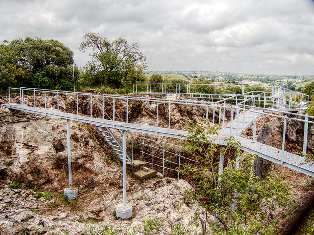
The entrance to the archaeological site at Sterkfontein, Cradle of Humankind (South Africa).
Ancient DNA of Humans
In 2008, a piece of a finger bone and a molar from a Siberian Cave were found that differed slightly from that of modern humans. The cave, called Denisova Cave, maintains an average temperature of 0ºC year round and was suspected to contain viable soft tissue. Bones in this cave were discovered that had similarities to modern humans and Neandertals. An initial mitochondrial DNA analysis revealed that these beings represented a distinct line of humans that overlapped with them in time (Krause et al., 2010). Analysis of the full nuclear genome followed and indicated that interbreeding existed between these Denisovans, Neandertals and modern humans (Reich et al., 2010). Furthermore, analysis of DNA from a 400,000 year old femur in Spain revealed that these three lines diverged from the species Homo heidelbergensis and that Denisovans were closest in sequence (Meyer et al., 2016).
Between modern humans, markers found in the mtDNA can be used to trace the migrations and origins along the maternal line. Similarly, VNTRs found on the Y chromosome have revealed migration patterns along paternal lines within men. Other markers, like the insertion points of transposable elements can be used to further describe the genetics and inheritance of modern humans while providing a snapshot into evolutionary history.
Yunis JJ, Prakash O. The origin of man: a chromosomal pictorial legacy. Science. 1982 Mar 19;215(4539):1525-30.
Chimpanzee Sequencing and Analysis Consortium. Initial sequence of the chimpanzee genome and comparison with the human genome. Nature. 2005 Sep 1;437(7055):69-87.
Krause, Johannes; Fu, Qiaomei; Good, Jeffrey M.; Viola, Bence; Shunkov, Michael V.; Derevianko, Anatoli P. & Pääbo, Svante (2010), “The complete mitochondrial DNA genome of an unknown hominin from southern Siberia”, Nature, 464 (7290): 894–897, doi:10.1038/nature08976, PMID 20336068
Reich, David; Green, Richard E.; Kircher, Martin; Krause, Johannes; Patterson, Nick; Durand, Eric Y.; Viola, Bence; Briggs, Adrian W. & Stenzel, Udo (2010), “Genetic history of an archaic hominin group from Denisova Cave in Siberia”, Nature, 468 (7327): 1053–1060, doi:10.1038/nature09710, PMID 21179161
Chromosomes are made of double stranded DNA molecules wound about histones and condensed into the familiar X-shape. Under regular functioning, these chromosomes are decondensed in the nucleus and not recognizable.
Chromosomes in Interphase are not visible individually. In preparation for nuclear division (mitosis or meiosis), they begin to organize tighter and condense in preparation for movement to subsequent daughter nuclei. The animation below illustrates the process of histone packaging and the molecular visualization of DNA replication. Histones are proteins that aid in packaging of the chromosomes into organized coils that give rise to the recognizable chromosomes during metaphase.
Large-scale genomic rearrangements result in genetic abnormalities. Biologists utilize a technique called a chromosome spread followed by a karyotype or karyogram. To make a chromosome spread, one blocks the progression of mitosis at metaphase where chromosomes are condensed into the structures we are familiar. A karyotype analysis is an arrangement of the chromosome spread into the homologous pairs of chromosomes.
A “spectral” karyotype of a female nucleus. Each homologous pair is “painted” to differentiate them.
Events associated with the improper separation of chromosomes during metaphase results in an alteration of chromosome number in the subsequent generation of cells. Using the Pop-beads, we can understand better how the timing of these events will lead to differences in the karyotype.


.jpg)


.jpg)


![By NIH, User:Phrood, En rouge [Public domain or Public domain], via Wikimedia Commons](https://openlab.citytech.cuny.edu/bio1-oer/files/2015/06/chromosomes.png)


