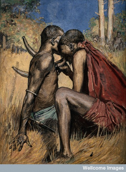
V0015962 An African medicine man or shaman applying the technique of
Credit: Wellcome Library, London. Wellcome Images
images@wellcome.ac.uk
http://wellcomeimages.org
An African medicine man or shaman applying the technique of cupping to a patient (using animal horns), which involves drawing blood to the surface of the body. Watercolour.
Published: –
Copyrighted work available under Creative Commons Attribution only licence CC BY 4.0 http://creativecommons.org/licenses/by/4.0/
This image depicts an African medicine man using the practice of cupping on a patient. This painting speaks to me because this is an ancient healing art that has transcended in to current Western use. Cupping is largely practiced in Chinese medicine, but it has dated back as early as 3000BC. It is believed to promote blood flow to heal muscle tension or pain in various areas.
Through heat or suction the skin is vacummed into a cup placed over the targeted area. In the image the healer is using what look to be a hollowed bone or horn of some sort and manually drawing the skin into it with his mouth. In modern use a rubber pump may be used in addition to silicone cups. The cups are then left in place for a short time before removing.
This is more of an alternative treatment to various ailments, so there is not a lot of research on its effectiveness