Learning Outcomes
• Explain the functions of the respiratory system
• Differentiate upper respiratory tract from lower respiratory tract
• List structures of the upper and lower respiratory tract
• Label a diagram of the respiratory tract from the nasal passages to the alveoli.
• List and identify major parts of the larynx and bronchial tree
Introduction
The respiratory system is involved the sense of smell, and the production of sound during speech. However, the main function of the respiratory system is in gas exchange. Gas exchange can be at the level of the lungs (lungs and blood), external respiration or between blood and tissue, internal respiration. In general respiration has to do with oxygen being taken up into the lungs and tissues, while carbon dioxide is expelled from the lungs as it makes its way to the lungs from cells and tissues. Animals and many other organisms are dependent on oxygen for survival. This oxygen is required as the final electron acceptor during cellular respiration, the key process involved in making ATP available to do work. During cellular respiration oxygen is utilized during the making of ATPs and carbon dioxide is released as a byproduct. The respiratory system begins with the nostrils and ends at the level of the alveoli. The respiratory system is made up of several structures divided into upper and lower respiratory system. The upper receives and conducts air to the lower respiratory tract. The lower respiratory conducts the incoming air to the alveoli in the lungs where gas exchange occurs.
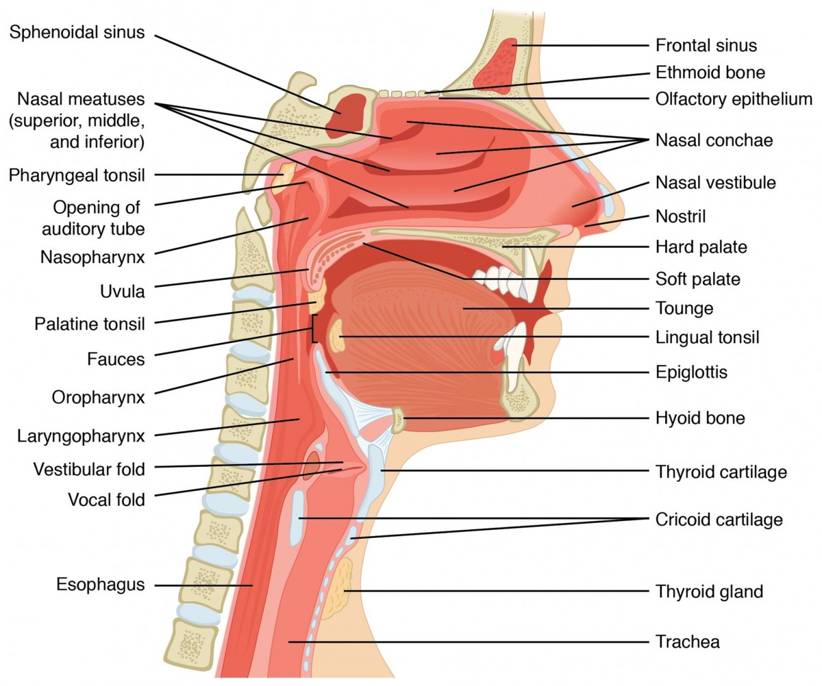
Figure 1. Upper respiratory tract. (Credit: OpenStax Anatomy and Physiology CC BY 3.0)
Upper Respiratory
External respiration begins with the nose, which houses a cavity, the nasal cavity, where air enters, warmed, moistened and cleaned. Externally the nose is made up of nasal bone and hyaline cartilage. External features include ala, apex and the dorsum nasi. The cavity is divided by the nasal septum giving rise to two sections, left and right. Each section as three bony structures, superior, middle and inferior conchae, on the lateral wall of the septum called conchae. These bony structures help increase the surface area. Between each concha is a meatus, superior, middle and inferior meatuses. Entrance to the cavity is through the nostrils or anterior nares. Each cavity has a roof, a floor, a lateral wall and a medial wall.Air enters the cavity and will eventually exit via the posterior or internal nares to the nasopharynx. Other features of the nasal cavity include sebaceous glands and mucous forming cells, hair follicles to filter the air, and olfactory cells involved in detecting odors. From the nasopharynx air makes it way to the rest of the pharynx, oropharynx and laryngopharynx, and enters the larynx.
The larynx controls air entering and leaving the lower respiratory tract. The larynx connects the pharynx, laryngopharynx to be more specific, to the trachea, the beginning of the lower respiratory tract. The larynx is composed of nine different cartilages; one pair of arytenoid cartilage, one pair of corniculate cartilages, one pair of cuneiform cartilages, a single thyroid cartilage, a single epiglottis and one cricoid cartilage. Except the epiglottis, all other cartilages are made up of hyaline cartilage. These cartilages serve as attachment sites for several muscles and ligaments, and also for protection. The two most inferior cartilages are the thyroid and cricoid cartilage. The thyroid is the largest of all the laryngeal cartilages. In most men the Adam’s apple or laryn
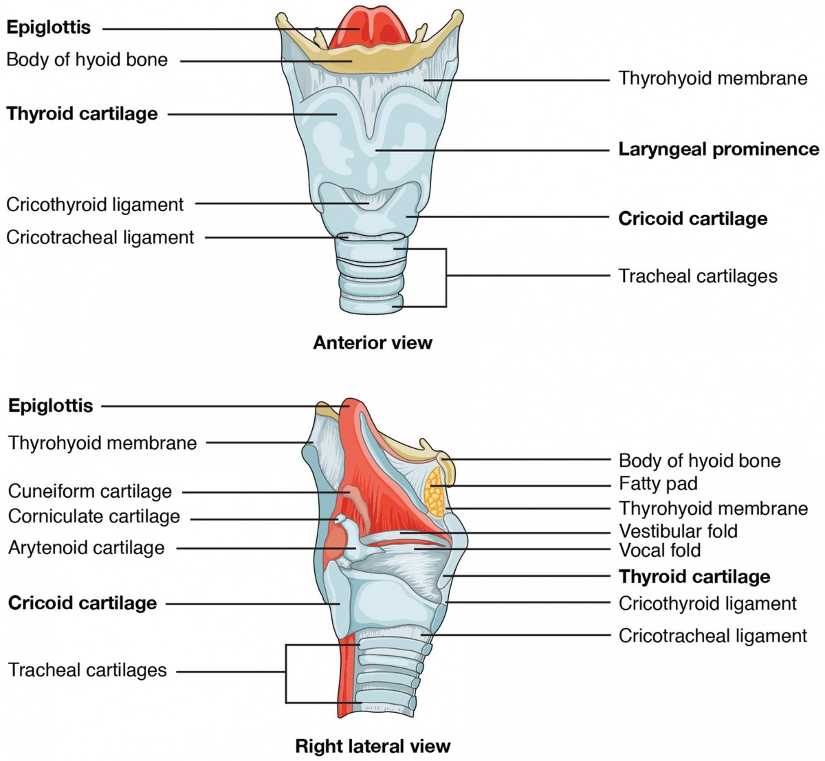
Figure 2. The larynx (Credit: OpenStax Anatomy and Physiology CC BY 3.0)
geal prominence, is the a protruding part of this cartilage. The cricoid cartilage is the only complete ringed cartilage of the larynx. The epiglottis is a flap of tissue made up of elastic cartilage which serves as a protective barrier for opening of the larynx, the glottis. The laryngopharyx is a common passage way for both food and air. When food is swallowed the epiglottis covers the glottis preventing food from entering. When there is no food and air is taken in, the epiglottis remains open and air gets through the glottis down to the lower respiratory tract. The glottis, a slit-like opening, is lined by two membranous folds forming true vocal cords
Lower Respiratory Tract
The lower respiratory tract begins with the trachea or the windpipe and end at the carina, where the trachea bifurcates into left and right primary bronchi. The trachea is a long tube, 10 – 13 cm, made up of 18 to 22 c-shaped hyaline cartilage connected posteriorly by trachealis muscle. The trachea bifurcates at the level of T4 giving rise to primary bronchi. These then enter each lung at the hilum forming subsequently branches of smaller tubes; secondary, tertiary, and bronchiole. Bronchioles the smallest branches of this bronchial tree, can be divided into terminal bronchioles and respiratory bronchioles. Respiratory bronchioles connect to several alveoli through alveolar ducts. Gas exchange occurs at the level of the alveoli. These tiny sacs are made up of three types of cells: type I alveolar cells, type II alveolar cells and alveolar macrophages. Most of the alveolar sac is made up of type I alveolar cell. Type II alveorlar cells are involved in producing pulmonary surfactant, a substance that reduce the surface tension of the alveoli. Alveolar macrophages, like other macrophages, are involved in removal of unwanted material and pathogens. Each alveolus is surrounded by several capillaries. Gas exchange takes place between the alveolar and capillaries via the respiratory membrane formed by the membrane of these two structures. Although this membrane is made up of three layers, the epithelial cells, the fused basement membrane and the capillary epithelium, it is thin enough to allow gas exchange.
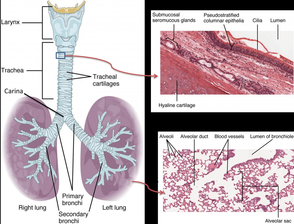
Figure 3. Lower respiratory tract and larynx. (Credit: OpenStax Anatomy and Physiology CC BY 3.0)
The lungs, left and right, consist of millions of alveolar sacs, between 250 – 800 million, organized as lobules. The right lung has three lobes while the left lung has two lobes. Each lobe is separated by fissures. In the case of the right lung there are the horizontal and oblique fissures giving rise to superior, middle and inferior lobe. The left lung has an oblique fissure giving rise to superior and inferior lobe. The left lung also has an indentation that accommodates part of the heart.

Figure 6. Lower respiratory tract. (Credit: OpenStax Anatomy and Physiology CC BY 3.0)

Figure 7. Lungs and pleura (Credit: OpenStax Anatomy and Physiology CC BY 3.0)
The lungs are both enclosed by a serous membrane called The pleura. This is a double layer membrane with one layer lining the thoracic cavity, parietal pleura and the other layer covering the lungs, the visceral pleura. Between the two layers is the pleural cavity, potential space with a thin film of fluid that allows the lungs to move freely with little friction. Air (pneumothorax), blood (hemothorax), and other fluids can accumulate in this cavity and lead to collapse of the lung(s). The pleura also help to keep the lungs in place and protect it from contact with other structures and organs. Because the parietal pleura is attached to thoracic wall, expansion of the thorax during inhalation also expands the lungs.
Cells of the Respiratory tract
The respiratory tract, from the nasal cavity to the alveoli, is composed of a variety of cells. The two main types are epithelial, eleven types, and mesenchymal, two types. The respiratory epithelium, from nose to the bronchi, is made up of mostly ciliated pseudostratified columnar epithelium, except the oro- and laryngopharynx which are made up of stratified squamous epithelium. In the alveoli the main cells, type I and II pnuemocytes, are squamous epithelium. Along the tract are goblet cells that secret mucous, club or clara cells found in bronchioles; they secret material which play protective role in the bronchioles.
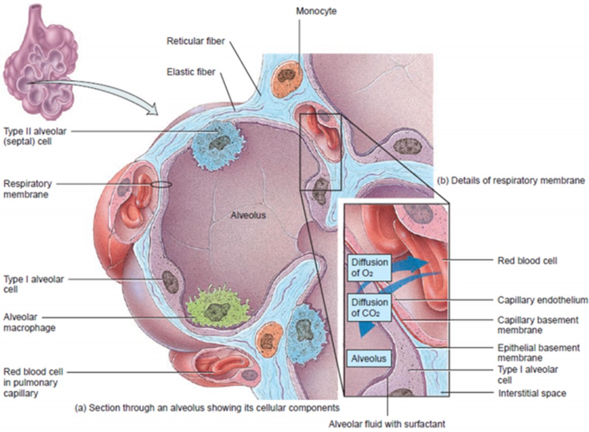
Figure 8. Cedit: Olympaid Student Center Wiki. Kokonilly CC BY 3.0

