
Human Reproductive System
The reproductive system is one of the systems that is, although with many similarities, strikingly different in males and females. At the early stages of development it is difficult to visually differentiate one from the other. The sex organs are not yet differentiated so hence no different organs exist.
These exercises will familiarize you with both male and female reproductive organs and structures.
Activity 1 – Male Reproductive System
")
Figure 1. Male reproductive system (Credit OpenStax Anatomy and Physiology CC BY 3.0).
Both male and female sex organs include gonads, duct systems, accessory organs and external genitalia. In males the gonad is the testis (pl. testes) or testicle. One of the primary roles of the testes is to produce and nourish sperm (mature sperm are called spermatozoa). Sperm more accurately refer to male sex cells compared to semen which include sperm, fluid and other factors produced and released by the accessory glands. Another primary role is to produce the hormone testosterone which is key to male sex differentiation.

Figure 2. Male anatomy (Picture from class model) (Credit Dr. Alcendor CC BY 4.0)
1. On any one of the models, identify the testis. It is housed in a sac called the scrotum. Identify the scrotum. What is the shape of the scrotum? ___________________ Within the scrotum are two layers of muscles, Dartos muscle and cremasteric muscle. Use your notes to name these two layers beginning with the layer closer to the skin of the scrotum (hint – DC). ____________________________________. The dartos muscle wrinkles the the scrotal sac while the cremasteric muscle moves the scrotal sac closer to the body.
2. Each of the two testes are covered by two layers, tunica vaginalis and tunica albuginea. The tunical albuginea is a layer of connective tissue while the tunica vaginalis is a layer of serous membrane. If you touch the testis on your model you are touching the tunica vaginalis, the visceral part of the tunica vaginalis. The parietal part of the tunica vaginalis is attached to the inside layer of the scrotum. Identify the two layers of the tunica vaginalis. What is between these two layers? Think of the pericardium or the pleural membrane. ____________________________
3. Within each testis are a number of tubules called the seminiferous tubules. It is within these tubules sperm are produced. From these seminiferous tubules sperm travel to the Rete testis then the efferent ductules on their way to the epididymis. Sperm will be stored in this structure until they are ready to be release during ejaculation. Identify the epididymis. How is it connected to each testis (top, bottom, middle, etc.)? ____________________________
4. Sperm travel from the epididymis to the vas deferens also called the ductus deferens. Identify the head, body and tail of the epididymis from your notes. In what direction would sperm travel on their way to the vas deferens? _________________________
5. Sperm will travel via the vas deferens to the ampulla of the vas deferens to the ejaculatory duct. The vas deferens will travel, along with blood vessels and nerves from the scrotum to the ampulla of the vas deferens via the spermatic cord. Identify the spermatic cord and follow it as it travels around the bladder on its way to the ampulla. Does it pass below or above the ureter? __________________________ What other major structures are found in the spermatic cord? _____________________________________________________________________________
6. From the ampulla, sperm travel to the ejaculatory duct and then to the first part of the urethra. In males the urethra is long and has three different segments. The first is the prostatic, the second the membranous and the third the spongy urethra. Which of these can you identify on your model? _______________________________
7. Identify the prostate, one of the accessory glands of the male reproductive system. Which organ or structure is located superiorly? ________________________________
8. The prostate produces and releases a milky fluid that will help activate and nourish the sperm on their way out during ejaculation. Describe the shape of the prostate. ________________________________________________________________________
9. Within the prostate there are two structures with openings within in; the opening of the bladder and the opening of the ejaculatory duct. The opening of the ejaculatory duct allows sperm and fluid from another accessory gland the seminal vesicle. The seminal vesicles are paired and provide fructose to energize the sperm as they travel. Identify the seminal vesicles. Describe the shape. What is the most obvious difference between the prostate gland and the seminal vesicle? _____________________
10. Another pair of accessory glands is the bulbourethral glands. These release fluid that will lubricate and help to clean urine from the urethra. The fluid is release into the spongy urethra as sperm travels out of the penis. It may be difficult to identify the bulbourethral glands on these models. From your notes, what is the difference in size between the three accessory glands? _______________________________________________________
11. Identify the penis. This is considered to be part of the male external genitalia. The other structures making up the external genitalia are the spongy urethra and the scrotum. Why are they called external genitalia? _____________________________________

Figure 4. Male Anatomy showing the erectile tissues in the penis. (Credit CC BY 4.0)
12. Examine the structure of the penis. Identify the erectile tissues, corpus cavernosum and corpus spongiosum. How are they different from each other? Into which of these does the spongy urethra travels? _________________________________________________________________________
13. The main region of the penis is the shaft. The root is the region closest to the bulbourethral glands. The third region is the glans penis which is the enlarged tip of the penis. Which erectile tissues continues to form the glans penis? ____________________________________
14. The end of the penis is the glands penis. This is the most sensitive part of the penis. It is usually covered by the prepuce or foreskin which serves to protect and also lubricate the glans penis. Which part of the male external genitalia is removed during circumcisions? ________________________________________________________________
15. Semen, which is sperm plus accessory fluid, having arrived in the spongy urethra will exit the penis via the ___________________________.
Questions
- Trace the path sperm travel from where they are produced to where they exit the penis.
- List the accessory glands and each give the number of glands present and the function each.
- List and give the function of the internal and external structures of the male reproductive system.
Activity 2 – Female reproductive system

Figure 5. Female reproductive system. (Credit OpenStax CC BY 3.0)
The female reproductive system is also consisting of gonads, ducts and accessory glands. The primary functions of the ovaries are (1) produce the female gametes or oocytes and (2) produce hormones such as estrogen and progesterone.
1. On a female model identify the ovaries. Like the testes, the ovaries produce and nourish sex cells or oocytes. Oocytes develop from primordial cells to mature oocytes and then are released into the infundibulum, the first part of the duct to deliver mature oocytes to the uterus. Identify the ovary or ovaries on your model. Describe the shape of an ovary. ________________________________________________________________
Compare the shape of an ovary with the shape of a testis. ________________________________________________________________
2. Identify the infundibulum, the part of the fallopian tube into which oocytes will enter on their way to the uterus. What hair-like structures are connected to each infundibulum? __________________________________________________________________________
3. Identify the rest of the fallopian tube as it makes its way to the uterus. It is in the fallopian tube that fertilization of the egg will take place. Which part of the male reproductive system does the fallopian tube have similar function to? _________________________________________________________________________
4. Where does the fallopian tube join the uterus? ______________________
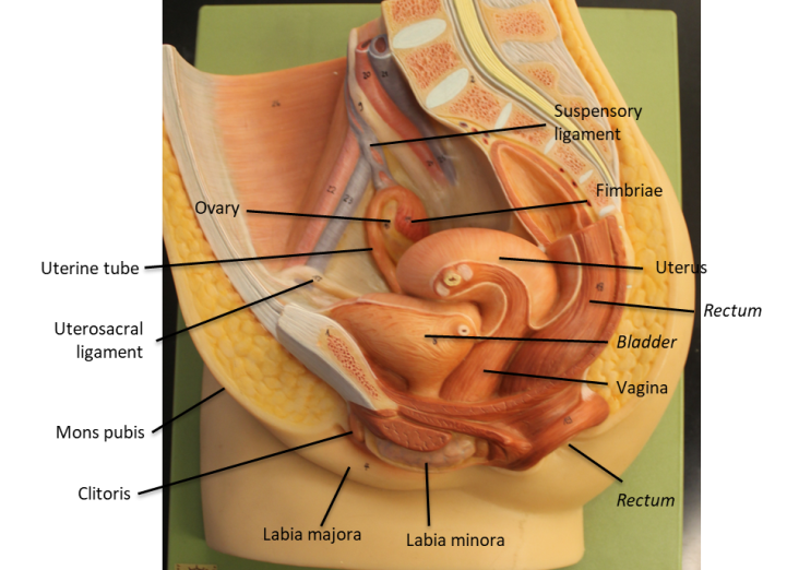
Figure 6. Female reproductive system using class model. Credit Dr. Alcendor

Figure 7. Female reproductive system using class model.
5. Identify the uterus, the thick muscular organ which house and protect the developing fetus. Describe its location in terms of the bladder._________________________________________
6. The uterus has four main regions: the fundus, the part superior to the opening of the fallopian tubes connect; the body, from the top to the region to the lower part of the uterus; the isthmus which leads into the last region called the cervix. Identify the four regions. Where does the fallopian tube connect to the uterus? _______________________________ Which region will receive sperm first? __________________________ The opening of the cervix is called external os.
7. Observe the wall of the uterus. It is made up of three layers: the inner layer (next to the opening) is the endometrium; the middle layer the myometrium and the outer layer the perimetrium. Based on your notes, which layer is thickest? _____________________ Which layer will be released during menstruation? _______________________
8. The structure connected inferiorly to the cervix is the vagina. This is where sperm will be released after ejaculation. The vagina is a muscular structure involved in passage of sperm, during menses and childbirth, BUT HAS NOTHING TO DO WITH PASSAGE OF URINE. Compare the thickness of the vagina with that of the uterus.
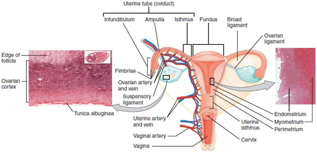
Figure 8. Female reproductive system internal organs and structures.
9. The opening of the vagina, vaginal orifice, is within part of the vestibule of the vulva, the region making up the external genitalia of females. Observe the distance between the vaginal orifice in the vestibule and the opening of the anus. How is it different from that of males? __________________________________________________________________________
10. Identify the urethra, which is not part of the reproductive system of females, and compare it to the vaginal canal. How are they different?
_____________________________________________________________________________
11. The vestibule is made up by two pairs of skin folds called the labia. The labia major is the larger pair and enclose the labia minor the smaller pair. How large are the labia major in reference to labia minora? ________________________________________________
12. Identify the clitoris found eternally and superior to the urethra. This is similar to the glans penis in males. It also contains erectile tissue called corpora cavernosa and is hooded by a prepuce. Like males it is the most sensitive part of the external genitalia.=fds
Question
1. Trace the path oocytes travel from where they are located to where they exit the vagina during menstruation.
2. List and give the function of the internal and external structures of the male reproductive system.

