Anatomy of the Heart – PDF
Student Learning Outcomes
- List the functions of the heart, heart structures and vessels.
- Identify the various parts of the human heart on charts and models.
- Describe the position of the heart in the thoracic cage.
- Identify major vessels entering and leaving the heart.
- Describe the structure and organization of cardiac muscles.
- Trace the pathway for blood through the heart via the pulmonary and systemic circuit.
- Dissect and identify the different parts, valves, chambers, etc. of the sheep heart.
Introduction
The heart plays one of the most vital functions of the body. It circulates blood all over the body carrying much needed material such as nutrients, hormones, proteins and other molecules, and oxygen while removing waste material and byproduct such as carbon dioxide, to be eliminated from excretory organs. The human cardiovascular system, like other mammals, is a closed system of heart and blood vessels. The heart pumps blood low in oxygen to the lungs, the pulmonary circuit, and pumps blood high in oxygen to the entire body, systemic circuit. The heart can be considered to be one of the most efficient pumps capable of enormous amount of pumping action. It contracts more than 100, 000 times in a day, almost 40 million times in a year, pumping about 5.25 liters of blood per minute. A typical adult’s heart is a little larger than the size of an adult’s closed fist weighing between 200-450 g. In certain diseased states, the heart can weight up to 1000 g.
Location of the Heart
The heart is located in the thoracic cavity occupying a space in the mediastinum; the area between the lungs. It lies between the second and the fifth intercostal spaces. The area of the heart where the large vessels (pulmonary trunk, aorta and vena cava) connect to the heart is called the base of the heart. The inferior tip of the heart is the apex. The apex, which is an extension of the left ventricle, lies to the left of the sternum at the level of the fourth and fifth ribs.
In the thoracic cage, the heart is protected, in addition to the pericardium, by bony ribs, cartilage from sternum and ribs, skin, fat, muscles and membranes. The heart itself is enclosed by a sac, called the pericardium or pericardial sac. This sac is made up of a top fibrous layer called the fibrous pericardium, and a serous membrane layer, serous pericardium. The pericardium is very tough, due particularly to the fibrous layer. Attached to that fibrous pericardium is the parietal pericardium, one of the layers of the two layers of the serous pericardium. The next layer of the serous pericardium, the visceral pericardium or epicardium, is attached to the heart. These layers are physically separated leaving a serous fluid-filled cavity called the pericardial cavity. This cavity is particularly important for allowing the expansion and contraction of the heart during its pumping action.
The fluid within the cavity acts as a lubricant reducing friction between the two layers. When one touches the heart, it is the visceral pericardium or epicardium that one touches. This layer is attached to the heart and is quiet difficult to separate. Extra fluid can accumulate in the pericardial cavity and prevent the heart from function. This condition is called cardiac tamponade.

Figure 1: Location of the heart in the thoracic cage. Credit: OpenStax Anatomy and Physiology CC BY 3.0
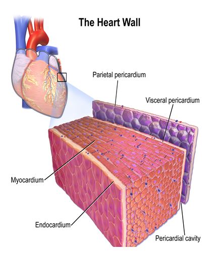
Figure 2: Layers of the myocardium. Credit: BruceBlaus. CC BY 3.0, ttps://commons. wikimedia.org/w/index.php ?curid=28761820
External Anatomy of the heart
Important structures on the external surface of the heart can be seen when the pericardium is retracted or the heart is outside the pericardium. Towards the base of the heart the auricles of the left and right atria are visible anteriorly. These are extensions of each atrium. At the base are the large vessels, the pulmonary trunk, the aorta and the superior and inferior vena cava. At the base of the heart are also major coronary arteries and cardiac veins. The left ventricle, the part of the heart where the apex is located, is separated from the right ventricle anteriorly by the anterior interventricular sulcus and posteriorly by the posterior interventricular sulcus. Within these sulci are coronary arteries and cardiac veins. Whereas coronary arteries receive blood from the base of the ascending aorta, blood from cardiac veins, except the anterior cardiac vein, empties into the coronary sinus, a thin-walled vein which empties into the right atrium. Coronary arteries, because of several factors such as diet, lack of exercise or other medical conditions, can narrow as a result of plaque or completely blocked leading to myocardial infarction or heart attack. Two other pairs of veins associated with the heart are the left and right pulmonary veins. These return blood from the lungs to the left ventricle. As discussed above, the outer layer of the heart is called the epicardium (visceral pericardium). It is attached to the myocardium, the thickest part of the heart muscle wall. On the inner side of the heart the endocardium, a layer of simple squamous epithelium, is attached to the myocardium. The thick myocardium forms twists and turn around both the atria and ventricles.

Anterior View

Posterior View Figure 3: Anterior and posterior views of the external surface of the heart.( Credit: OpenStax Anatomy and Physiology) CC BY 3.0
Activity 1 – Heart Location and Eternal Structures
Internal Structures of the Heart
The heart is a pump with four chambers, two atria and two ventricles. The atria are located superiorly and are the receiving chambers of the heart, returning blood to the heart from all over the body. The right atrium receives blood from the superior and inferior vena cava, while the left atrium receives blood from the lungs via the pulmonary veins. The superior vena cava receives blood from parts of the body above the diaphragm, while the inferior vena cava receives blood from below the diaphragm. The atria are separated from each other by the interatrial septum, and from the ventricles by the tricuspid (three cusps) valve on the right and the bicuspid (two cusps) valve on the left. Although both atria are similar in structure, the right atrium has more pronounced muscular ridges called pectinate muscles. These help the RA dilate during adverse loading conditions. Additionally, the remnant of a fetal structure, foramen ovale, is visible as the fossa ovalis. The foramen ovale allowed most fetal blood to bypass the right ventricle and the pulmonary circulation and instead enter the left atrium on its way to the aorta. The two ventricles are separated by a longer and thicker interventricular septum. The left and right ventricles have marked differences (see table below). The most obvious difference is the thicker left ventricle muscular wall. The left ventricle pumps blood to the entire body, therefore the pressure is greater in the left ventricle. Like the atria there are irregular muscular ridges or projections in the ventricles called trabeculae carneae. They help strengthen the heart walls and assist the papillary muscles. Both ventricles have papillary muscles, small muscles extending from the ventricular walls. The right ventricle has three papillary muscles attached to cusps of the tricuspid valve, while the left has two papillary muscles attached to the cusps of the bicuspid or mitral valve. Papillary muscle is attached to on cusp of the atrioventricular valves by chordae tendineae or heart strings, and prevent valves from over extending under ventricular pressure.
The flow of blood follows a one way direction. Blood is prevented from back flow as a results valves. Between the atria and the ventricles are atrioventricular valves (AV); tricuspid valves on the right and bicuspid or mitral valve on the left. Blood leaving the right ventricle is prevented from returning into the ventricle by the pulmonary semilunar valves, while blood leaving the right ventricle is controlled by the aortic semilunar valve. These valves are anchored or held in place by a dense layer of connective tissue called the cardiac skeleton or fibrous skeleton of the heart.

Figure 4: Internal structures of the heart. (Credit: OpenStax Anatomy and Physiology CC BY 3.0)
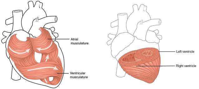
Figure 5: Muscular organization of the heart (credit: Openstax Anatomy and Physiology CC BY 3.0)
The flow of blood in the heart follows a one way direction primarily due to the four valves. Between the atria and the ventricles are atrioventricular valves (AV); tricuspid valves (three cusps are present) on the right and bicuspid or mitral valve (two cusps are present) on the left. These prevent blood from flowing back into the atrium when the ventricles are contracting. Blood leaving the right ventricle is prevented from returning into the ventricle by the pulmonary semilunar valves, while blood leaving the right ventricle is controlled by the aortic semilunar valve. Both semilunar valves are made up of three cusps. These valves are anchored or held in place by a dense layer of connective tissue called the cardiac skeleton or fibrous skeleton of the heart.


Activity 2 – Internal Structures of the Heart
Blood Flow through the heart
The heat receives oxygen poor blood returning from the systemic circuit from upper and lower part of the body, the superior vena cava and inferior vena cava respectively. Blood from these large vessels empty into the right atrium and will eventually flow into the right ventricle via the tricuspid valve. From the right ventricle blood flows through the pulmonary semilunar valve into the pulmonary trunk on its way to the pulmonary arteries and to the lungs. In the lungs oxygen diffuses into the blood while carbon dioxide diffuses out of the blood into the lungs on its way out of the body. The oxygenated blood returns to the left atrium via the pulmonary veins. From the left atrium blood flows through the mitral or bicuspid valve into the left ventricle. Eventually the left ventricle will pump the blood through the opening of the aortic semilunar valve into the aorta where it will be distributed all over the body. The circulation of blood to and from the lungs involves the pulmonary circuit, while to and from the rest of the body is the systemic circuit. Besides the pulmonary circuit and systemic circuit, blood also flow through what is called coronary circulation. The heart muscles and tissues receives blood through left and right coronary arteries originating from the base of the first part of the aorta, the ascending aorta. From there it flows to major coronary arteries; the anterior interventricular artery, the circumflex artery, marginal arteries and posterior interventricular artery. Blood leaving the heart muscles and tissues is drained by coronary veins. The major ones are the great cardiac vein following the interventricular sulcus, the posterior cardiac vein, the middle cardiac vein and small cardiac veins. Blood will eventually flow into the coronary sinus on its way to the right atrium.

Figure 6: Blood flow through the heart. (Credit: OpenStax Anatomy and Physiology CC BY 3.0)
Diseases and disorders affecting the heart
- Congenital heart diseases – problem or defect in heart and heart structure that someone is born with. Example atrial septal defect, where there is an opening between the right and left atria, or ventricular septal defect where there is a hole in the interventricular septum between the right and left ventricles.
- Coronary artery disease – the main coronary arteries become block as a result of byildup of plaque or atherosclerosis.
- Cardiomyopathy – Disease of the heart muscle. The heart muscle may be thicker or larger than normal.
- Pericarditis – Inflammation of the pericardium.
- Arrhythmia – Problems with the rhythm of the heart. The heart may be beating too fast, tachycardia, or too slow, bradycardia.
- Angina – discomfort or chest pain.
- Heart attack (myocardial infarction) – occurs when the heart does not receive blood as a result of blockage of coronary arteries.
- Heart failure – heart is unable to pump sufficient blood to the body.
- Aortic aneurysms – enlarging or bulging of the aorta
- Valvular heart disease – problems with proper functioning of the four heart valves.

