Contents
Size-exclusion of dye molecules
As a demonstration, the instructor may illustrate the concept of size exclusion on a set of mixed food coloring.

- Pack column with 3 ml sepharose slurry
- Let the column empty over a beaker
- Carefully load 0.2 ml of food coloring mixture onto the column
- Place 10 tubes on a rack under the column
- Place 1 ml buffer on column and collect 0.5 ml fractions
- Continue to add buffer 1 ml at a time until all fractions have been collected
Size-exclusion of Proteins
This exercise seeks to purify Green Fluorescent Protein (GFP) or Blue Fluorescent Protein (BFP) from bacterial lysate. These proteins have a specific size of 238 amino acids and are 40,000 daltons (40kD). Based on their specific size, they will have a specific rate of migration through the size exclusion resin. Remember that the bacterial lysate is full of additional proteins that are not your protein of interest that we are attempting to isolate.
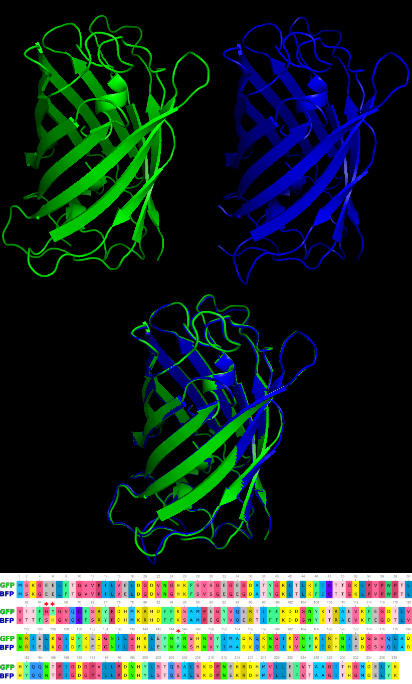
Drops of fluid will be collected in fractions. The fractions containing the fluorescent proteins will be found only in specific fractions that will be visible under UV illumination.
- Vertically mount the column on a ring stand. Make sure it is straight.
- Slide the cap onto the spout at the bottom of the column.
- Mix the slurry (molecular sieve) thoroughly by swirling or gently stirring.
- Carefully pipet 2 ml of the mixed slurry into the column by letting it stream down the inside walls of the column.
- Place an empty beaker under the column to collect wash buffer.
- Remove the cap from the bottom of the column and allow the matrix to pack into the column.
- Label eight microcentrifuge tubes #1-8.
- Slowly load the column with 0.2ml of the GFP extract. Allow the extract to completely enter the column.
- Add 1ml of elution buffer on top of resin without disturbing the resin
-
- Add buffer slowly (several drops at a time) to avoid diluting the protein sample.
- Using the graduated marks on the sides of the tubes, collect 0.5ml fractions in the labeled microcentrifuge tubes.
- Continue to add 1ml buffer and collect fractions until all tubes are full
- Check all fractions by using long wave U.V. light to identify tubes that contain the fluorescent GFP or BFP proteins.
- Further purification may be performed with a different resin with the few fractions containing the protein of interest
- Protein samples should be run on an acrylamide gel and stained against all proteins to check the purity of the sample or fluorescence measurements taken
Tags: analysis

