Category Archives: Uncategorized

Homework #1

The image I chose from Wellcome Images does not have an artist’s name but it was “taken from the Apocalypsis S. Johannis cum glossis et Vita S. Johannis; Ars Moriendi (circa 1420 to 1430), which were gynaecological texts that included information about conception, pregnancy, and childbirth”.  This image is an ink and watercolor illustration that shows a caesarean section in medieval times. The woman on the table is still cut open, bleeding and dead. The nurse is holding the baby that was just born in a blanket. The surgeon, although doesn’t look like a typical surgeon is holding a knife, a knife that you would cut a piece of meat with. This picture is not easy to look at, especially for a woman. Most women, who underwent a C-section in medieval times, did not make it due to the severe bleeding that occurred. Sterile techniques were not in place, allowing infection to play a big role in death. This image relates to the images we saw in class with the woman and child at the doctor’s office. Similar to those images, we can conclude that back then doctor offices did not look sterile, proper or how we would expect them to look. Physicians looked like ordinary people.  As you can see in this drawing, the room has no surgical supplies besides the butcher knife. There is also no means of sedation or pain medication. The amount of pain, infections and blood loss experienced by the mother was more than enough to guarantee death after the birth of a child.
Homework #1

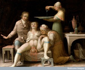
The image I chose is an oil painting of a child birthing scene by a French painter, which depicts a very detailed picture of a woman in the 1800 giving birth.

In this painting, the woman giving birth has a very pale, distressed facial expression and it seems as though she has lost a lot of hair on the crown of her head which is a symptom of pregnancy. She is also undressed from the stomach down but is still wearing socks with a blue band to prevent the sock from falling. The man behind her might be her husband holding her up. He seems more excited and happy rather then sharing the distress the woman is in because of the position of his hand on the side. There are two other woman that seem to be helping the woman with the child birth. One of them is obviously much older than anyone in the entire painting kneeling down, calmly and professionally wrapping the pregnant woman’s leg while holding the body of the infant. The other woman is standing behind the whole scene pouring water on a pan for the infant to lie on to bathe him/her. From the furniture and utensils in the painting, the family doesn’t seem to be very wealthy and the room seems fairly empty with not much furniture or objects.

What is so striking about this painting to me is that the painter chose to show the full frontal view of what giving birth was like at that time including the blood and half of the infant’s body sticking out of the woman’s vaginal region. The entire vaginal area is very much blatantly displayed with blood dripping from the cloth to the floor. Compared to the images seen in class, this painting is sending a more positive vibe. Child birth may seem very painful and bloody but it’s actually a very happy event to give life.


V0017247 A birth-scene. Oil painting by a French (?) painter, Ă…bo, Sw Credit: Wellcome Library, London. Wellcome Images images@wellcome.ac.uk http://wellcomeimages.org A birth-scene. Oil painting by a French (?) painter, Ă…bo, Sweden (later Turku, Finland), 1800. 1800 Published:  -  Copyrighted work available under Creative Commons Attribution only licence CC BY 4.0 http://creativecommons.org/licenses/by/4.0/

V0017247 A birth-scene. Oil painting by a French (?) painter, Ă…bo, Sw
Credit: Wellcome Library, London. Wellcome Images
images@wellcome.ac.uk
http://wellcomeimages.org
A birth-scene. Oil painting by a French (?) painter, Ă…bo, Sweden (later Turku, Finland), 1800.
1800 Published: –
Copyrighted work available under Creative Commons Attribution only licence CC BY 4.0 http://creativecommons.org/licenses/by/4.0/

Homework #1

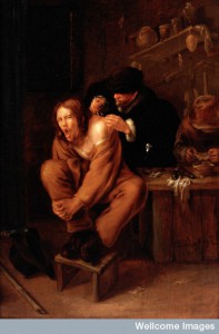
L0027284 A surgeon applying medicine to a wound in the shoulder of a Credit: Wellcome Library, London. Wellcome Images images@wellcome.ac.uk http://wellcomeimages.org A surgeon applying medicine to a wound in the shoulder of a man in pain. Oil painting by Gerrit Lundens, 1649. Oil 1649 By: Gerrit LundensPublished: 1649 Copyrighted work available under Creative Commons Attribution only licence CC BY 4.0 http://creativecommons.org/licenses/by/4.0/

L0027284 A surgeon applying medicine to a wound in the shoulder of a
Credit: Wellcome Library, London. Wellcome Images
images@wellcome.ac.uk
http://wellcomeimages.org
A surgeon applying medicine to a wound in the shoulder of a man in pain. Oil painting by Gerrit Lundens, 1649.
Oil
1649 By: Gerrit LundensPublished: 1649
Copyrighted work available under Creative Commons Attribution only licence CC BY 4.0 http://creativecommons.org/licenses/by/4.0/

The image I picked from the Wellcome Database is called “A surgeon applying medicine to a wound in the shoulder of a man in pain.” by Gerrit Lundens. It captured my attention because of the face of the man from the painting. There is three people on the painting. One doctor, one patient, and one look to be the assistant who is preparing the medicines. The patient is sitting on the table while the doctor is putting medicine on his wound on his shoulder. The patient is wearing an overall and a pair of work boots, therefore I think he might got his shoulder hurt at work. However, His face tells us that the medicine is giving him more pain, which is a normal phenomenon because the medicine is killing the bacteria, meanwhile, the skin gets more sensitive to the stimulus from the environment when it’s wounded. It reminds me of a image we saw in class, which was about the woman is having a headache and the doctor is checking on her. In both images, I can find the origin of the distress by examining the details of the image carefully; Also, both of their face expressions give us a generally idea of what is happening there.

Homework 1

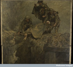
L0050213 World War I: stretcher bearers of the Royal Army Medical Credit: Wellcome Library, London. Wellcome Images images@wellcome.ac.uk http://wellcomeimages.org World War I: stretcher bearers of the Royal Army Medical Corps (RAMC) lifting a wounded man out of a trench. Painting by Gilbert Rogers. Below, a trench in which a man stands lifting one end of a stretcher over the top of the trench. Above, three man receive the stretcher, on which lies a man with his face covered with a bloody bandage. Left, a shell explodes. The background is buff, the trench and the figures are predominantly grey, yellow and green, giving an impression of mud everywhere.  Of the men in the upper half, two have armbands with red crosses on a white ground, in addition to uniform badges with red crosses on, while the third, on the left, has only the red cross badge on his uniform, and no armband.  The body of the man on the stretcher is foreshortened to show the soles of his boots, a device also found in paintings by Mantegna, Borgianni, and Rembrandt. 1919 By: Gilbert RogersPublished:  -  Copyrighted work available under Creative Commons Attribution only licence CC BY 4.0 http://creativecommons.org/licenses/by/4.0/

L0050213 World War I: stretcher bearers of the Royal Army Medical
Credit: Wellcome Library, London. Wellcome Images
images@wellcome.ac.uk
http://wellcomeimages.org
World War I: stretcher bearers of the Royal Army Medical Corps (RAMC) lifting a wounded man out of a trench. Painting by Gilbert Rogers.
Below, a trench in which a man stands lifting one end of a stretcher over the top of the trench. Above, three man receive the stretcher, on which lies a man with his face covered with a bloody bandage. Left, a shell explodes. The background is buff, the trench and the figures are predominantly grey, yellow and green, giving an impression of mud everywhere.
Of the men in the upper half, two have armbands with red crosses on a white ground, in addition to uniform badges with red crosses on, while the third, on the left, has only the red cross badge on his uniform, and no armband.
The body of the man on the stretcher is foreshortened to show the soles of his boots, a device also found in paintings by Mantegna, Borgianni, and Rembrandt.
1919 By: Gilbert RogersPublished: –
Copyrighted work available under Creative Commons Attribution only licence CC BY 4.0 http://creativecommons.org/licenses/by/4.0/

The imagine I have chosen is, World War I: stretcher bearers of the Royal Army Medical. I chose this image because of how powerful it is to me, and also because most medical advances have been an achievement of warfare. Throughout history almost every medical advance has been something that has come forth because of human’s capacity to kill each other with the most creative ways that we can think of at the time. But with all the evil, and torment that are associated with war there has been some good that has come out of it. Almost every major medical breakthrough that has been achieved by human beings has come about because of war. Through ways to treat trauma injuries, mental disorders, and infections, the cures have come because physicians and governments are looking for ways to save their fellow citizens from certain ravages of war. From the first medic/shaman that first treated injured soldiers in ancient battle fields in the near Middle East, to the advancement that we have today. The military has been the testing grounds for certain procedures, theories, and innovations of already existing techniques and equipment. For as long as there has been warfare, there has been men who try to help the wounded men heal. Most of humanity has benefited from all the medical discoveries that have been made through battle fields’ hospitals and research to treat those that have come home with physical and mental wounds.

Wellcome Image

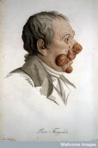
Wellcome Library offers a variety of images to choose from. I chose an image called Pian Fungoide by Jean Luis-Marie Alibert that was published in 1806 in Paris. Jean-Louis Alibert was a dermatologist born on 1768 in southern France and died in 1837. Pian Fungoide was a term used for the first time by him and the name came from the fact that this condition had skin lesions that resembled the mushrooms. This picture is a profile drawing of the face of a middle age man, that has large deep brown overgrowth of lesions on his face. The background is white and plain. The image does not say much about the class status of the patient. Hi does not have a clean haircut, and the seems to be wearing a white coat.  The name of the condition has been printed on the bottom of the paint, and I`m not sure if the name of the author has been printed on the left bottom side of the drawing. Initially when I saw this image, it captured my attention because I had no idea what the condition could be and was interested on exploring the image and condition further. The lesions on his face are very large and I can imagine the effect they had on patient himself. They make his face look scary especially considering that this drawing was from the early 1800. Since this condition caused such deformity, I feel that people might have been scared of him and even seen him as an evil human being. Even though we see the right side of his face, I noticed the growth is a little obvious left eyebrows, but we cant see the much of the other side. It seems as if the right side must have been more deformed than the left.

L0020973 J. L. M. Alibert, Description des maladies de la peau. Credit: Wellcome Library, London. Wellcome Images images@wellcome.ac.uk http://wellcomeimages.org Pian Fungoide Description des maladies de la peau observees l'Hopital Saint-Louis Jean Louis-Marie Alibert Published: 1806 Copyrighted work available under Creative Commons Attribution only licence CC BY 4.0 http://creativecommons.org/licenses/by/4.0/

L0020973 J. L. M. Alibert, Description des maladies de la peau.
Credit: Wellcome Library, London. Wellcome Images
images@wellcome.ac.uk
http://wellcomeimages.org
Pian Fungoide
Description des maladies de la peau observees l’Hopital Saint-Louis
Jean Louis-Marie Alibert
Published: 1806
Copyrighted work available under Creative Commons Attribution only licence CC BY 4.0 http://creativecommons.org/licenses/by/4.0/


Homework # 1 Wellcome Database

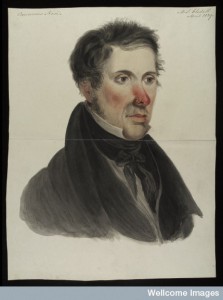
The image I chose from the Wellcome Library is called “Mr Gledell, suffering from a rodent disease which has.” This watercolour painting, painted in 1829, is one of the six portraits forming a group of works showing gentlefolk of Leeds with grievous illnesses. The painting shows a young, white man with short black hair dressed in a black coat and stock, possibly in upper class society. He is almost emotionless. It is hard to tell whether he is calm or unhappy. And the grayish white on the background of the painting gives a feeling of sadness. The painting captured my attention because of his red nose. It looks like the red nose that we all get in the cold winter season. However, looking closer to the painting we could find a flat red scaly patch on his right nostril.  Indeed, this young man suffers from a rodent disease which has consumed his left nostril. This young man is facing half left, so we could not see the disfigurement on left side of his nose resulted from the rodent disease. This is similar to the images we saw in class. They all depict some types of diseases, illnesses or cancers. The only difference in the painting is the hidden of the disfigurement because the young man is facing half left and looking away from the painter.

L0037317 Mr Gledell, suffering from a rodent disease which has Credit: Wellcome Library, London. Wellcome Images images@wellcome.ac.uk http://wellcomeimages.org Mr Gledell, suffering from a rodent disease which has consumed his left nostril. One of six portraits forming a group of works depicting gentlefolk of Leeds with grievous illnesses. A youngish man, facing half left, dressed in a black coat and stock, his left nostril eaten by a cancer and the whole nose a vivid red. Watercolour 1829 Published: April 1829. Copyrighted work available under Creative Commons Attribution only licence CC BY 4.0 http://creativecommons.org/licenses/by/4.0/

L0037317 Mr Gledell, suffering from a rodent disease which has
Credit: Wellcome Library, London. Wellcome Images
images@wellcome.ac.uk
http://wellcomeimages.org
Mr Gledell, suffering from a rodent disease which has consumed his left nostril. One of six portraits forming a group of works depicting gentlefolk of Leeds with grievous illnesses. A youngish man, facing half left, dressed in a black coat and stock, his left nostril eaten by a cancer and the whole nose a vivid red.
Watercolour
1829 Published: April 1829.
Copyrighted work available under Creative Commons Attribution only licence CC BY 4.0 http://creativecommons.org/licenses/by/4.0/