Contents [hide]
Reading
Chromosomal Basis of Inherited Disease
Learning Objectives
- Differentiate between histones and nucleosomes
- Define the terms karyotype
- Describe the differences in organisms with respect to chromsome numbers
- Define the abnormalities in chromosomal numbers: polyploidy, aneuploidy: trisomy and
- monosomy, and mosiacism and their causing mechanisms.
- Define the abnormalities in chromosomal structure: deletions, duplications, translocations, and inversions.
- Distinguish among the following “mistakes” in meiosis: non-disjunction, deletion, duplication, inversion, and translocation; give an example of each.
- Distinguish among Down’s syndrome, Klinefelter syndrome, and Turner syndrome.
- Know what amniocentesis is, and compare it to chorionic villus sampling.
- Distinguish between physiological therapy, protein therapy, and gene therapy.
Chromosome Packaging

Chromosomes are made of double stranded DNA molecules wound about histones and condensed into the familiar X-shape. Under regular functioning, these chromosomes are decondensed in the nucleus and not recognizable. Credit: KES47 [CC BY 3.0]

False color representation of chromosomes in a nucleus illustrating the 24 types of human chromosomes in their decondensed state. Credit: Andreas Bolzer, Gregor Kreth, Irina Solovei, Daniela Koehler, Kaan Saracoglu, Christine Fauth, Stefan Müller, Roland Eils, Christoph Cremer, Michael R. Speicher, Thomas Cremer [CC BY 2.5]
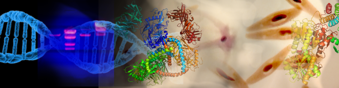
Chromosomes in Interphase are not visible individually and are loosely packaged within the nucleus. In preparation for nuclear division (mitosis or meiosis), they begin to organize tighter and condense in preparation for movement to subsequent daughter nuclei. The animation below illustrates the process of histone packaging and the molecular visualization of DNA replication. Histones are proteins that aid in packaging of the chromosomes into organized coils that give rise to the recognizable chromosomes during metaphase.

Credit: Richard Wheeler [CC-BY-SA-3.0]
DNA is negatively charged due to the Phosphate backbone. Histones are positively charged proteins that associate with eukaryotic DNA to spool and compact the DNA. The form into basic units called nucleosomes. The basic nucleosome core is composed of and octamer of histone proteins (H2A, H2B, H3 and H4).

Nucleosome structure illustrating the spooling of DNA around the core histone octamer. Credit: Richard Wheeler (Zephyris), Rekymanto [CC-BY-SA-3.0]
Chromosome Structure
The familiar X-shape of chromosomes can only be observed during cell division when there are 2 sister chromatids. These chromosomes come in different physical configurations.

The basic shapes of chromosomes and their structure. I: Telocentric – centromere placement very close to the top, p arms barely visible if visible at all II: Acrocentric – q arms are still much longer than the p arms, but the p arms are longer than it those in telocentric III: Submetacentric – p and q arms are very close in length but not equal IV: Metacentric – the p arm and the q arms are equal in length A: Short arm (p arm) B: Centromere C: Long arm (q arm) D: Sister Chromatid Credit: Fockey003 [CC BY-SA 4.0]

A “spectral” karyotype of a female nucleus. Each homologous pair is “painted” to differentiate them.
Events associated with the improper separation of chromosomes during metaphase results in an alteration of chromosome number in the subsequent generation of cells.

