Contents [hide]
Learning Objectives
- Describe the components and basic structure of the plasma membrane, and explain what is meant by the fluid-mosaic model.
- Explain how a cell membrane regulates interactions within environments.
- Differentiate between diffusion, osmosis, and dialysis.
- Describe the solute/solvent movements into and out of a cell under hypertonic, hypotonic, and iso-tonic conditions.
- Explain and give examples of generalized endocytosis and exocytosis
- Understand the importance of selective permeability in biological systems.
- Differentiate among diffusion, facilitated diffusion and active transport.
- Describe the operation of the Na+– K+ pump
- Compare and contrast cilia and flagella in terms of their structure and function.
- List and describe the various junctions, linkages, and connections that occur between cells.
- Understand the biochemistry of phospholipids and how they are organized into membranes.
Understanding Membranes
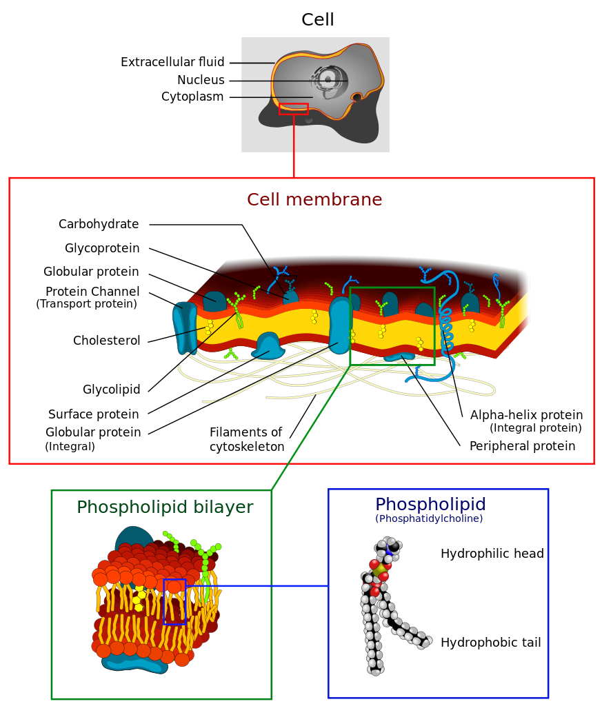 The cell membrane is the barrier that separates the cytoplasm from the external world. The cell membrane consists primarily of phospholipids in a bilayer. Phospholipids are amphipathic with a polar head (phosphate group) and a hydrophobic tail (2 hydrocarbon chains). Due to the chemical properties of the heads being attracted to water and the tails having a desire to avoid water, phospholipids self assemble into micelles. Cell membranes form from a phospholipid bilayer where the lipid tails interact with each other and the phosphate heads face the external water environment or the internal cytoplasm of the cell.
 The cell membrane does not solely consist of phospholipids but also have proteins and cholesterol inserted into the bilayer. As the image of the bilayer above indicates, the molecules are constantly moving and flow in a lateral motion. Cholesterol modulates the fluidity of this motion. Proteins associated with the membrane may sit on either side (peripheral proteins) of the membrane or pass through both layers of the membrane (transmembrane proteins). The model that describes the components of the cellular membrane is referred to as the Fluid Mosaic Model. This model states that the cell membrane is a mosaic of 1)Phospholipids 2)Proteins 3) cholesterol that move about in a side to side motion.

The fluid mosaic of phospholipids, proteins and cholesterol that create the selective barrier between the interior and the exterior of the cell.

Small uncharged molecules pass through the double layer of phospholipids. Polar, charged or large molecules have great difficulty passing through the membrane and require the aid of transmembrane proteins. An example of a transmembrane protein that facilitates movement of a polar substance is aquaporin, which permits the free movement of water.

 1) a single transmembrane α-helix (bitopic membrane protein). 2) a polytopic transmembrane α-helical protein. 3) a polytopic transmembrane β-sheet protein. The membrane is represented in light green.

A classic experiment demonstrates the fluidity of membranes through fusion of membrane vesicles. In this experiment, two vesicle donors are chemically labeled with different fluorescent dyes or antibodies. Through a chemical induction, the vesicles fuse together. Over time, the blending of the lipid components can be observed microscopically as the membrane contents diffuse into each other.


