Contents
Reading
- PDF (OpenStax)
- 4.1 Studying cells (OpenStax CNX)
- Microscopy (Khan Academy)
- 4.2 Prokaryotic Cells (OpenStax CNX)
- Prokaryotic Cell Structure (Khan Academy)
Learning Objectives
- State the components of the cell theory.
- Illustrate differences in methods of studying cells
- Describe the domains under Prokarya and differentiate them from Eukarya
- Give reasons to explain why cells are generally very small
- State the importance of multicellularity.
Slide Show
Discovery of cells
Hooke’s Cell

Illustration of Hooke’s microscope

The empty chambers of cork inspired Hooke to coin the term “Cell”
The father of Microbiology: van Leeuwenhoek

van Leeuwenhoek’s microscope
Though van Leeuwenhoek’s apparatus was simple, the magnifying power of his lenses and his curiosity enabled him to perform great scientific observations on the the microscopic world. He was ridiculed for fabricating his observations of protists at first. Ever the scientist, van Leeuwenhoek examined samples of his own diarrhea to discover Giardia intestinalis. While he did not make the connection of the causative nature of this microorganism, he described the details of the way this organism could propel itself through the medium in great detail.
van Leeuwenhoek also visualized sperm cells from animals and observed the process of fertilization of egg cells. This observation was extremely important because it negated the idea of spontaneous generation, the notion that life can spring forth from inanimate material. Spontaneous generation previously explained the observations that maggots could arise from decaying meat and fleas arose from dust.
.jpg)
Modern micrograph of Giardia intestinalis (Kingdom Protista) Credit: Doc. RNDr. Josef Reischig, CSc. [CC-BY-SA 3.0]
Life arises from life
The discovery of the microscopic world enlightened scientists and aided in abolishing spontaneous generation and furthered invented the field of microbiology. Prior to this, a popular idea for spread of diseases like bubonic plague involved the spread of miasma (“bad air”). To protect himself from the bad air, the plague doctor wore a beaked mask filled with fragrant items and a built in respirator.
Pasteur, fermentation and Germ theory of disease
Pasteur conducted experiments with the fermentation of nutrient broth in goose-necked flasks. Upon the discovery of the microscopic world, it was thought that microbes could be spread. Goose-necked flasks could be boiled to sterilize the nutrients while protecting the contents from contacting external microbes. the removal of the goose-neck or movement of broth into the goose-neck resulted in the subsequent fermentation of nutrient broth while the intact flasks remained sterile.

Animated version of Pasteur’s experiments on spontaneous generation Credit: Thebiologyprimer [CC-BY 4.0]

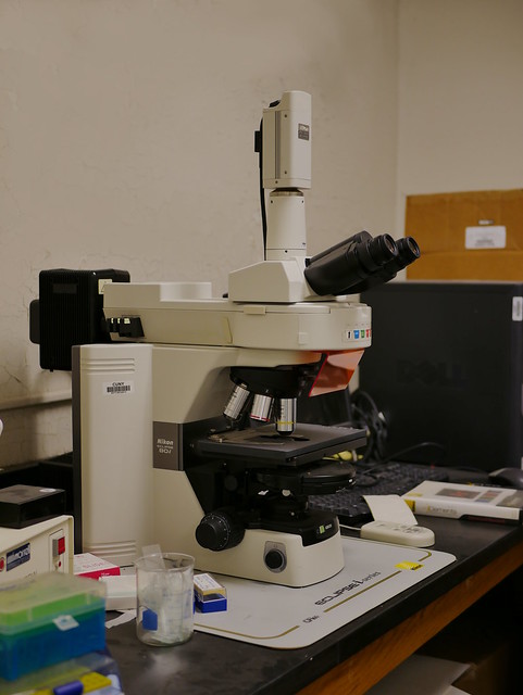
Modern Compound Microscope
Unlike van Leeuwenhoek’s single lens microscope, we now combine the magnifying power of multiple lenses in what is called a compound microscope.

Compound Light Microscope. Credit: jeremy Seto [CC-BY-NC-SA]
Electron Microscopy
An electron microscope is a microscope that uses a beam of accelerated electrons as a source of illumination. A scanning transmission electron microscope has magnifications of up to about 10,000,000x whereas most light microscopes have a limit of magnifications below 2000x
Transmission Electron Microsopy

Schematic of Transmitting Electron Microscope. Credit: Gringer [CC-BY-SA 3.0]
Scanning Electron Microscopy

Pollen under a scanning electron microscope. [CC0]
Fluorescence Microscopy
Fluorescent Microscope. Credit: jeremy Seto [CC-BY-NC-SA]

Schematic of a fluorescent micrscope. Credit: Henry Mühlpfordt [CC-BY-SA 3.0]
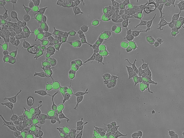
Light micrograph overlayed with a fluorescent micrograph of green fluorescent protein expressing cells. Credit: jeremy Seto [CC-BY-NC-SA]
Dissecting Microscope
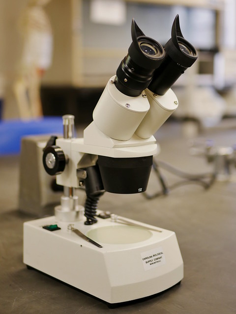
Dissecting Microscope. Credit: jeremy Seto [CC-BY-NC-SA]
Prokaryotes

Ring of life diagram illustrating the divergence of prokaryotes and the subsequent endosymbiosis that gave rise to eukaryotes. Credit: Maulucioni [CC-BY-SA 3.0]

Schematic diagram of a prokaryotic cell. Credit: Ali Zifan [CC-BY-SA 4.0]

Basic shapes of bacteria. [CC0]

