
You will find pictures of radiographs taken in clinic, pathology seen in clinic and interesting cases.
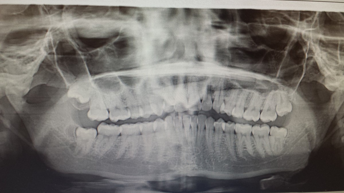
Panoramic of a 24years old patient showing 3rd molars that do not have enough space for full eruption.
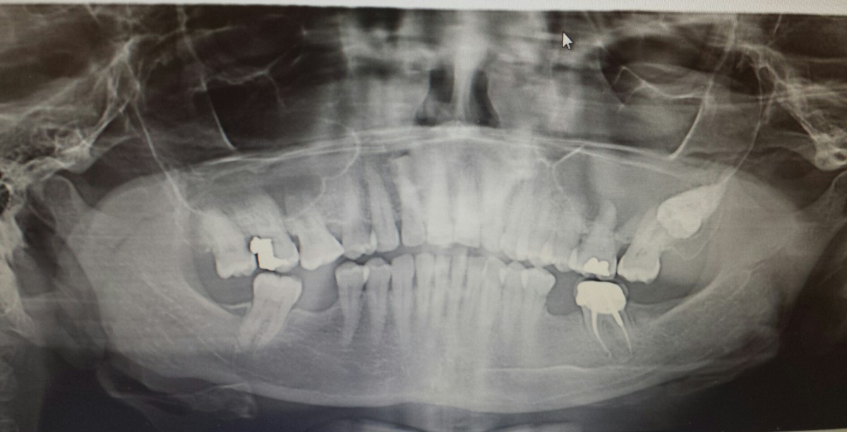
Panoramic of 52 years old patient with impacted #16; patient was never aware of the position of this tooth and this was the first time this patient had a panoramic taken.
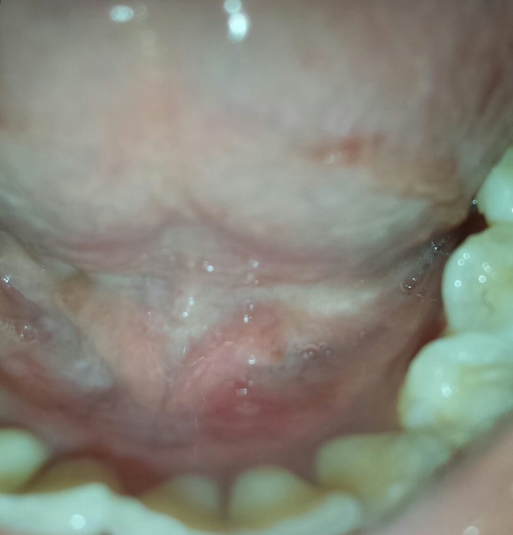
Apthous ulcer
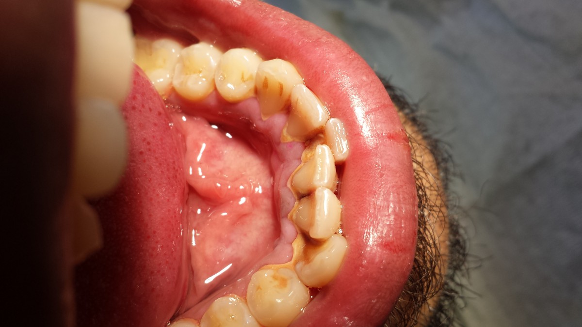
This patient presented with generalized heavy supragingival and subgingival calculus
After periodontal scaling and root planning

This patient was very motivated to get treatment done and was implementing OHI that was taught, patient was very happy when treatment was completed.
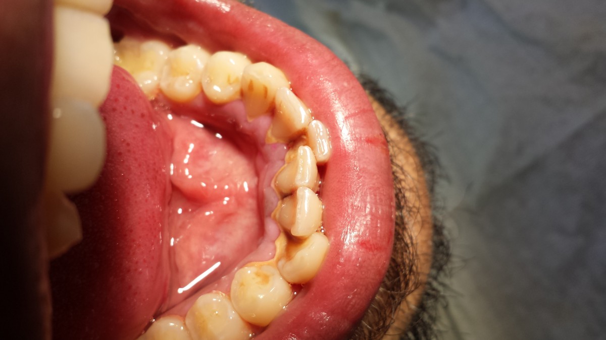
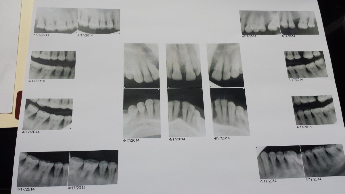
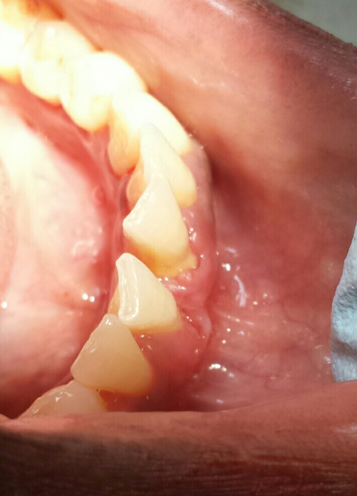
42 years old patient with Type III periodontal disease. Below is a copy of radiographs which was taken when patient went for a dental exam:
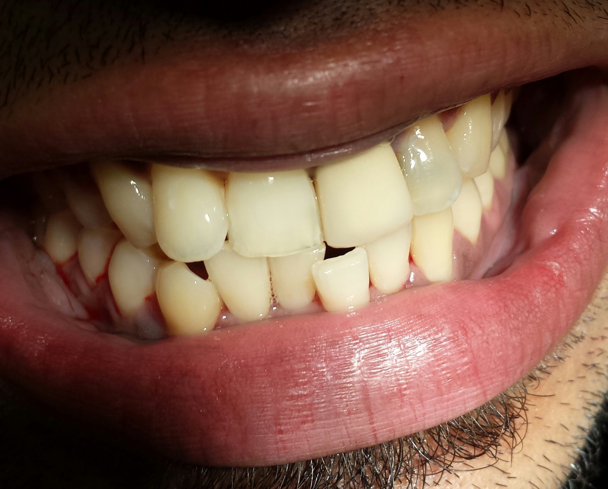
Mucogingival defect noted for the above patient (photo taken after UR and LR were scaled):
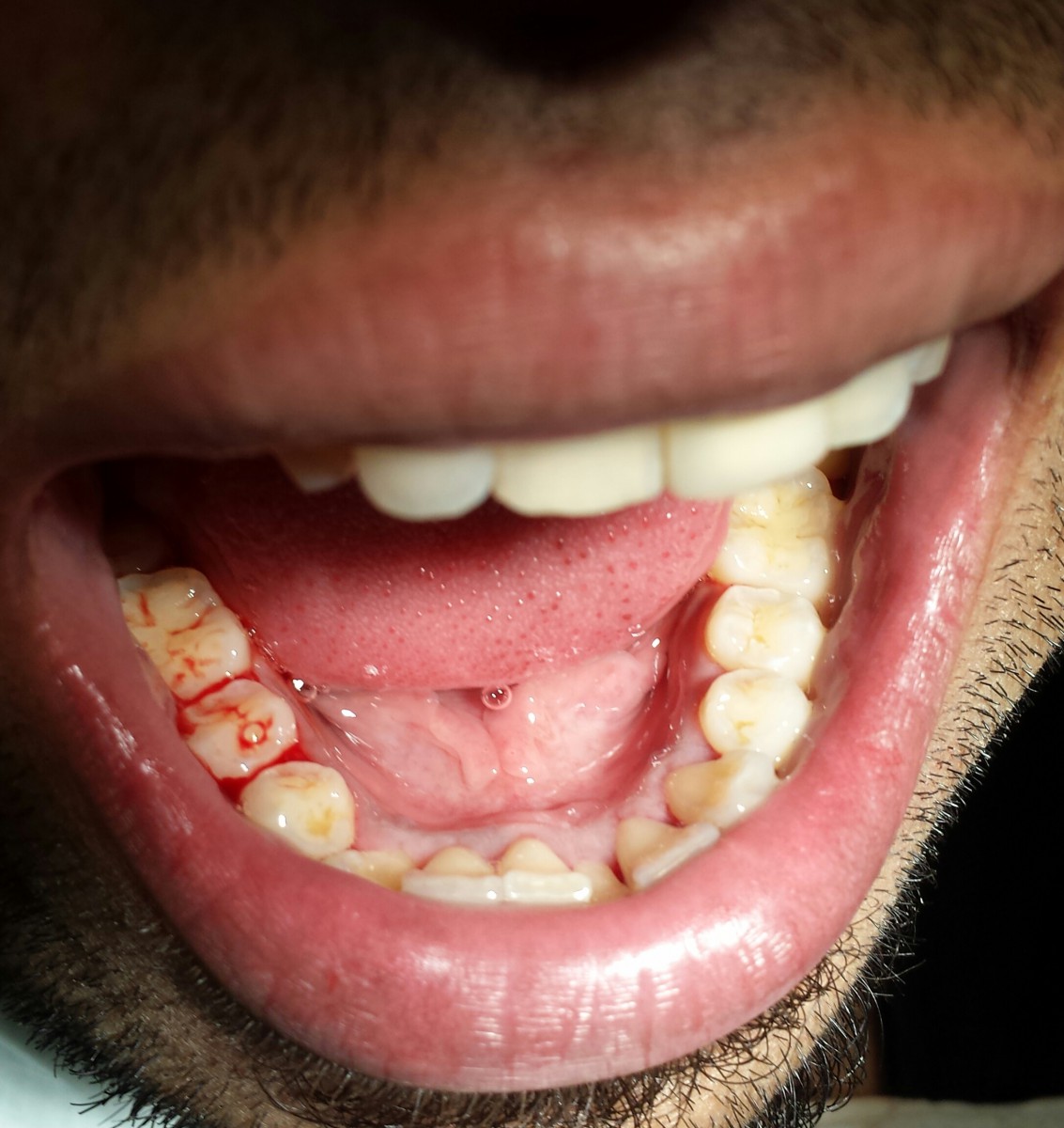
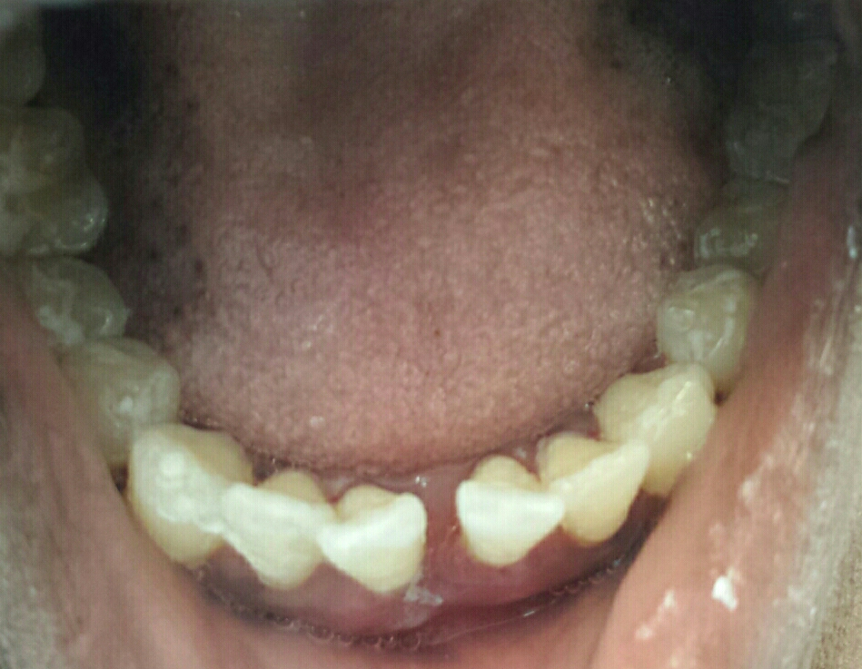
After periodontal scaling and root planing for the above patient (taken after fluoride varnish application):
Headstart Fluoride Varnish Program
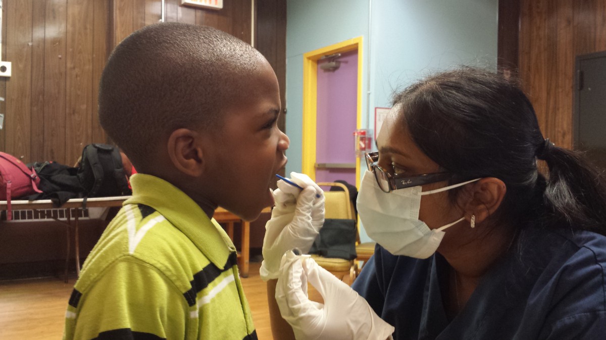
We went to Glenwood Center where we did screening for decay with a tongue depressor and illumination. We applied fluoride varnish after the screening. This was a great experience and it felt very good to be part of this public service.

