
6 month recare/H/Type II: 28 year old Hispanic male. Patient in overall good health. Not currently taking medications and/or vitamin supplements. Nonsmoker. BP: 121/79, Pulse: 69. ASA I. No cc.
Dentition: Amalgam restorations in posteriors. Maxillary and mandibular 3rd molars fully erupted/functional. Attrition: #6-#11 and #22-#27.
Periodontal: Marginal redness and inflammation in maxillary anterior region. Interdental papillae of mandibular anteriors is flaccid and appears lacerated due to patient’s reported attempts to floss. Attached gingiva of maxillary posteriors is spongy, rolled margins present. Generalized Type II due to 4-5mm probing depths, moderate BUP, and recession. Localized Type III due to 5-7mm probing depths, moderate BUP, recession, and radiographic evidence of moderate vertical bone loss in maxillary posteriors. Generalized heavy sub-gingival calculus and localized supra-gingival calculus on the lingual surfaces of the mandibular anteriors. Minimal staining.
Radiographs: 4 vertical bite-wings exposed to evaluate the height of the alveolar bone where 5-7mm probing depths were present. Radiographs revealed localized vertical bone loss in maxillary posteriors. No evidence of caries/PAP.
Oral Hygiene Instruction: Patient reports using a soft bristle toothbrush and floss-picks. Recommendations: Use of electric toothbrush 2x daily for 2 mins. due to insufficient plaque removal and calculus accumulation. Advised patient to maneuver floss-picks up and down against the teeth when flossing in order to accommodate for the curvature of the teeth, as opposed to placing the floss in and out. Recommended use of Crest Gum Detoxify dentifrice and Listerine Antiseptic mouthrinse- 20mL for 30 seconds daily.
Treatment Plan/Implementation:
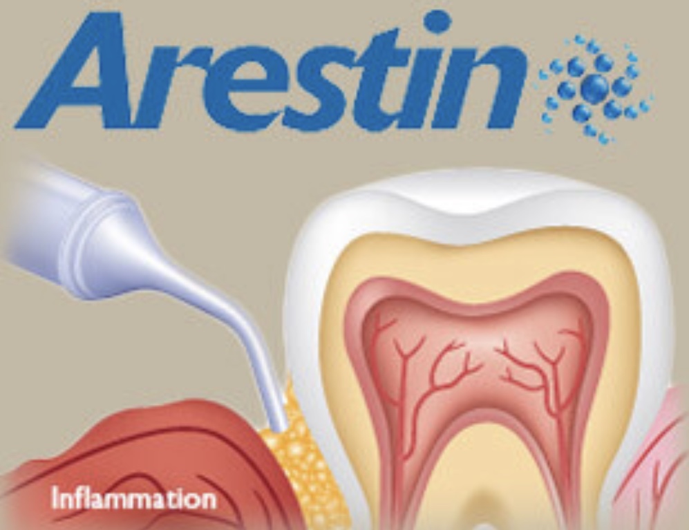
V1: Completed assessments. 4VBW exposed and interpreted. OHI. Handscaled and used ultrasonics on UL quad. Arestin to be placed next visit: #14-MB, #14-ML, #15-MB, and #15-ML.
V2: (1 week) Re-eval. UL quad. OHI: Review modified bass toothbrush method and flossing. Administer Arestin. Handscaled and used ultrasonics on LL, UR, and LR. 5% Sodium Fluoride Varnish applied; Post-operative instructions given verbally.
V3: (4 weeks) Arestin eval. Probe areas in which Arestin was placed. Document changes in appearance of gingival tissue and updated probing measurements.
1mm decrease in probing depths: #15- MB/ML
2mm decrease in probing depths: #14-MB/ML
Arestin treatment was confirmed successful. Gingival tissue appeared pink, firmly attached, smooth contour of marginal gingiva in maxillary posteriors. Decrease in probing depths due to Arestin. Patient was thoroughly surprised with the positive feedback and motivated to maintain measurements below 5 and 7mm.
N.V.: 3 month recare (August 2018)

