Clic to see Detailed Case Scenario 1
Written assignment done from a treated patient in clinic during the Fall 2016 semester.

Extra Oral Examination Identification of severe dry lip. Scabs and scar from chronically cracking lip. Patient was referred for evaluation to a dermatologist.

Identification of lesions when performing intraoral examination. Patient presents with aphthous ulcers on left side oral mucosa adjacent to tooth #21. Follow up appointment 2 weeks after lesions were resolved.
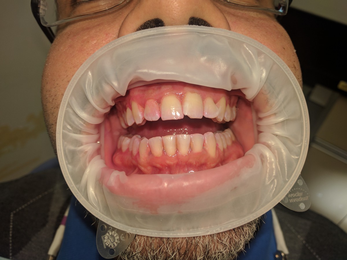
Pre Procedural Picture Heavy case value, Periodontal Type I. Disclosing agent reveals plaque mainly on interproximal areas and tooth #7.

Post Procedural Picture Scaling of 4 quadrants with ultrasonics and manual instruments.

Patient with history of HepC, Heavy case value, Periodontal Type III. This is a picture of his revisit appointment. Right side evaluation after manual instrumentation on previous appointment.
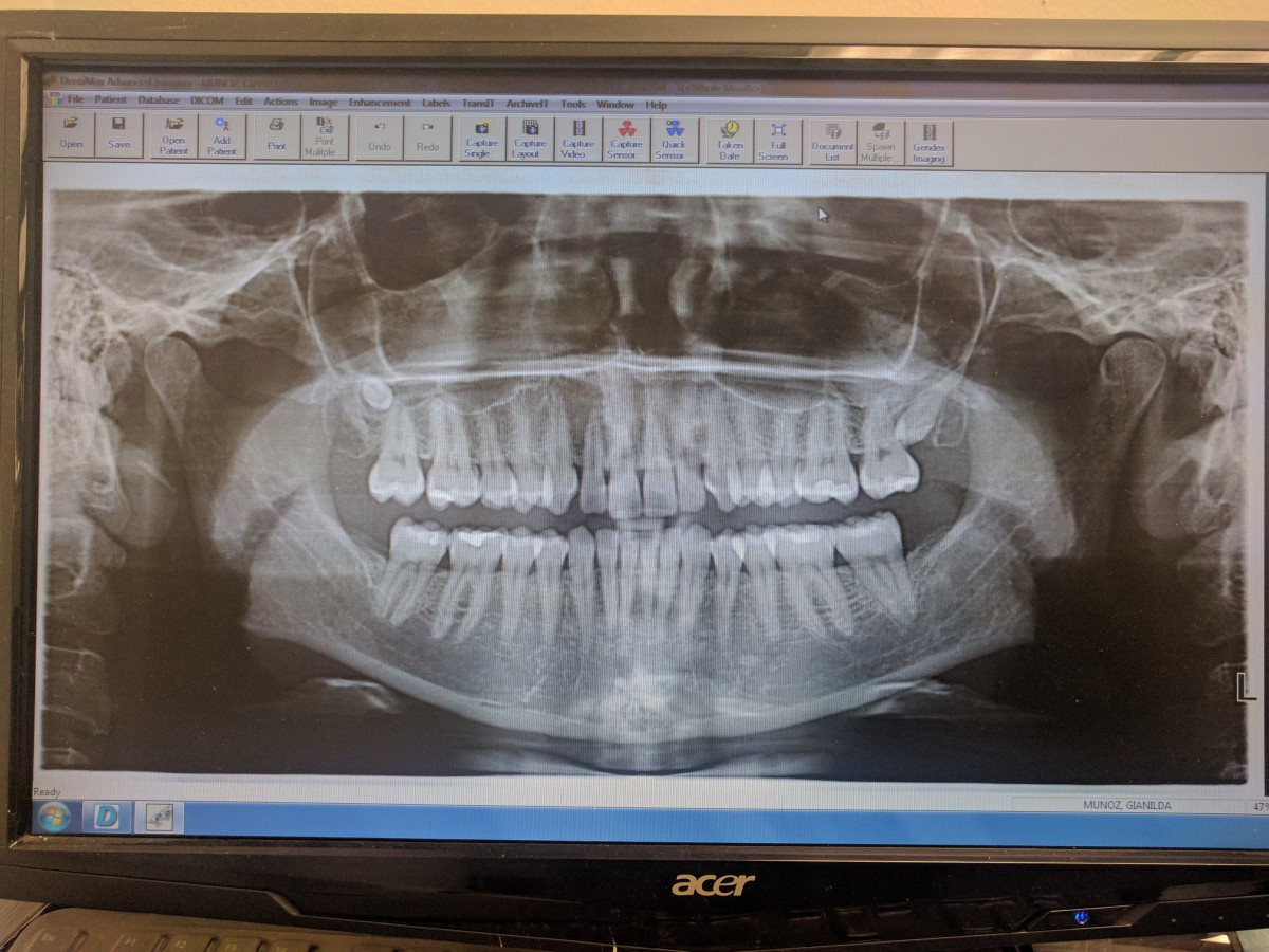
Panoramic taken on a patient congenitally missing lower third molars. Radiographic detection of upper impacted microdont third molar teeth.

