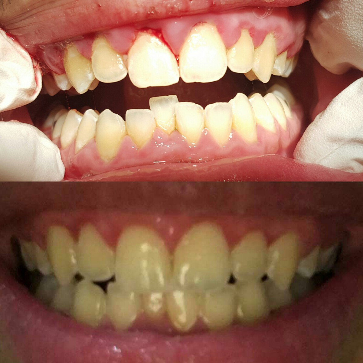
(Before: Top picture)
20 year old patient presents with severe edema and friable tissue, as well as, spontaneous bleeding. Pocket depths ranging from 3-7mm, no radiographic bone loss evident. Pocket depths are pseudo pockets due to extreme inflammation. Generalized heavy sub and supra gingival deposits. Referral given for patient to have a check up due to hemorrhagic bleeding during treatment, and up to two days following.
(4 weeks post op: Bottom picture) Poor picture quality due to picture being taken by patient and sent via email. The patient was unable to present for their 8 week perio evaluation, when the picture was set to be taken.
Gingival tissue displays generalized moderate marginal inflammation. Patient states they are not complying with recommended home care. Significant changes in tissue contour and consistency evident. Patient has yet to see a doctor for blood work. Spontaneous bleeding still occurring occasionally.
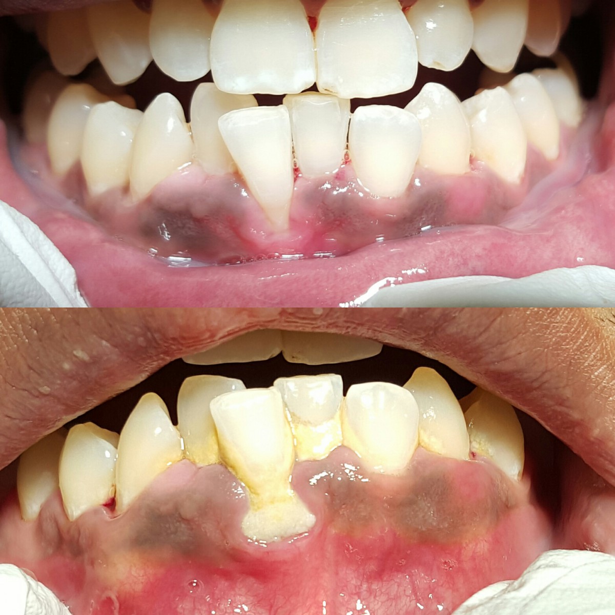
(Before: Bottom picture)
30 year old male in good health. Pocket depths ranging from 3-8mm. Approximately 10% bone loss. Severe halitosis. Tissue displays severe edema, and recession on #25.
(1 week Post op: Top picture)
Pocket depths decreased to 2-6mm. Halitosis resolved. Tissue is beginning to show improvement in consistency and contour. #25 has a mucogingival defect. Suggested a consult with both an orthodontist to correct bite, and periodontist for possible graft.
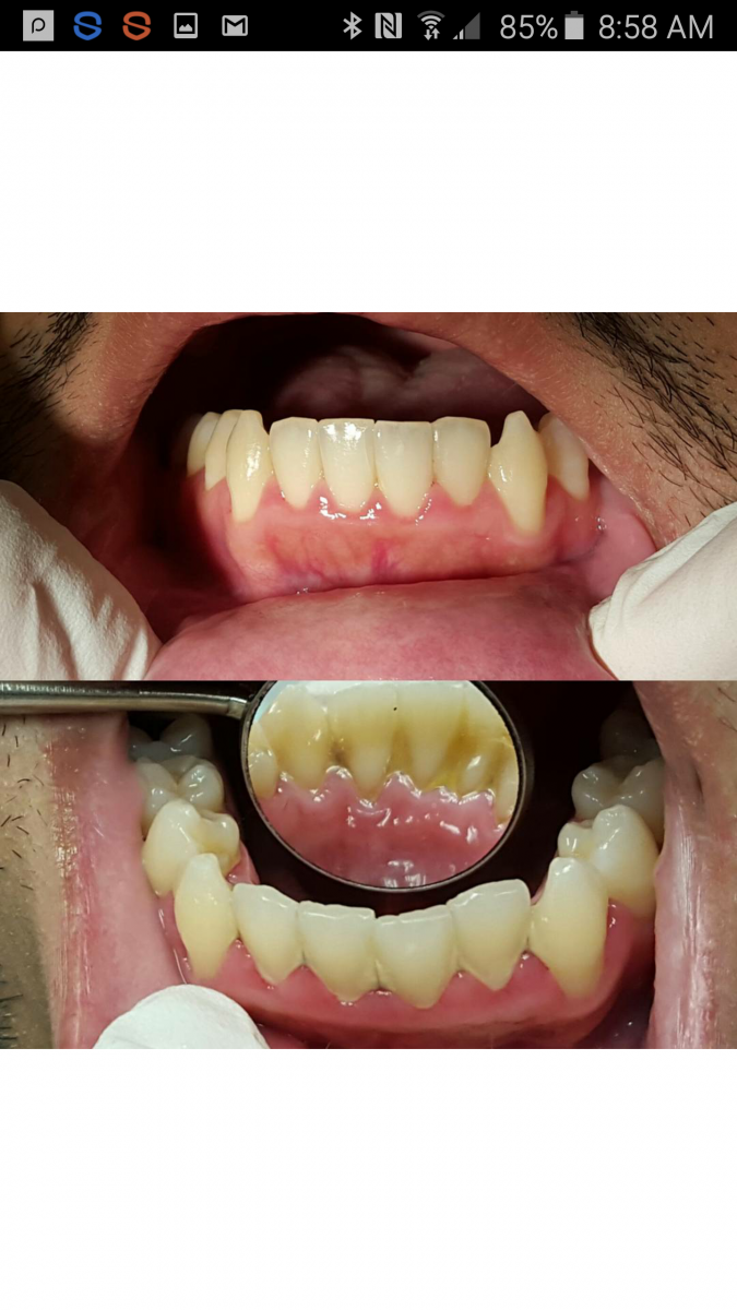
(Before: Bottom picture)
26 Year old male. Hemorrhagic bleeding. Pocket depths ranging from 4-8 mm. Tissue is flaccid and edematous.
(3 weeks post op: top picture)
Arestin placed in multiple sites. Pockets depths decreased to 3-5 mm pockets. Bleeding ceased. Tissue has significantly reduced inflammation, and tissue adhesion progressing.
RESEARCH


Hi Justine,
I was curious to see your portfolio.
Anyway, on case #01, it would be good to note that there is significant loss of attached gingiva on #25, resulting in a muccogingival defect. This pt. may benefit from a grafting procedure or even extraction. I wonder, is that tooth in crossbite and that contributed to exaggerated loss of attachment. Also, at week one, soft tissue is still healing. Compare this to case # 2 at week three, very nice, improved appearance of the tissue. Great results.
Thank you Dr. Noel. I have taken your advice and made some adjustments. I advised the patient to have a consult with an orthodontist, as well as a periodontist for graft. I think the attachment loss stems from the crowding. The patient does have a cross bite.