Table of Contents
Introductory video about membranes
The components of the membranes
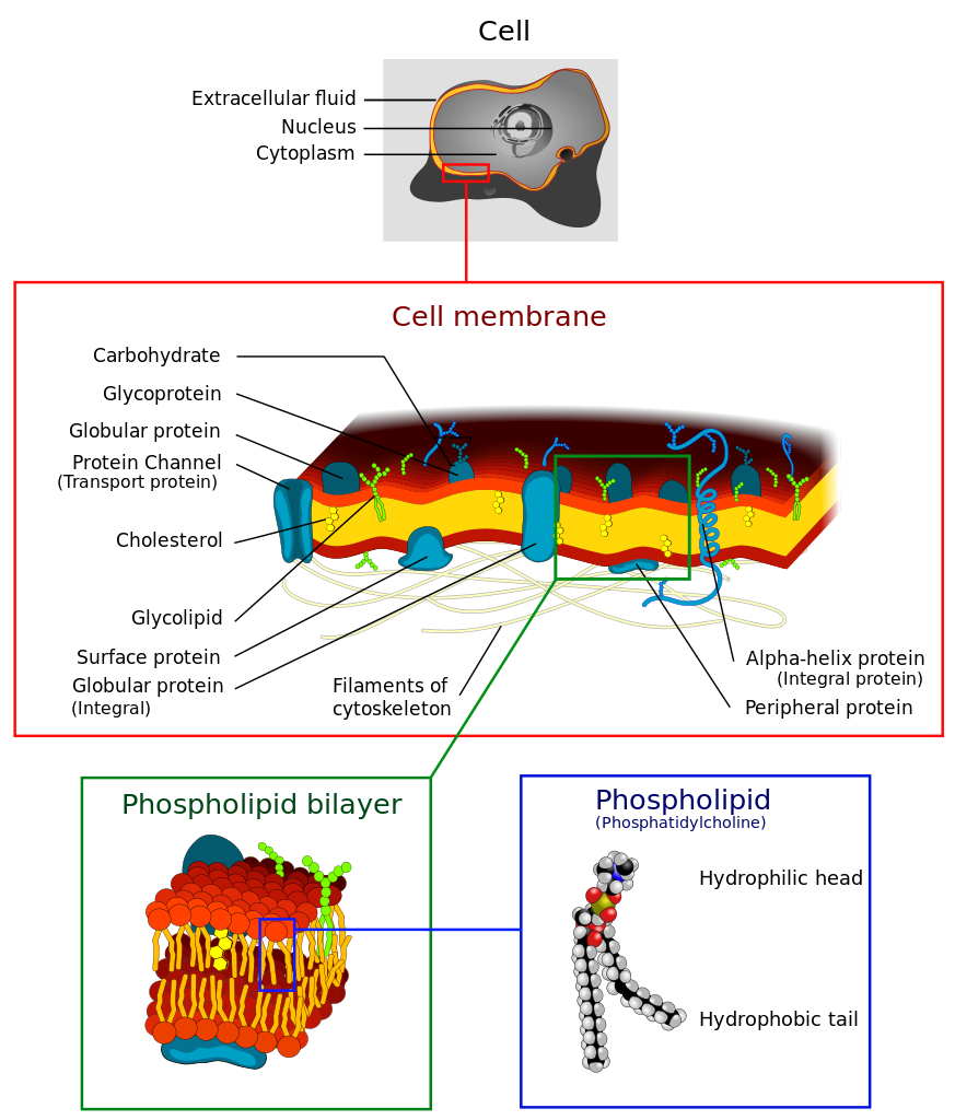


The cell membrane is the barrier that separates the cytoplasm from the external world. The cell membrane consists primarily of phospholipids in a bilayer. Phospholipids are amphipathic with a polar head (phosphate group) and a hydrophobic tail (2 hydrocarbon chains). Due to the chemical properties of the heads being attracted to water and the tails having a desire to avoid water, phospholipids self assemble into micelles. Cell membranes form from a phospholipid bilayer where the lipid tails interact with each other and the phosphate heads face the external water environment or the internal cytoplasm of the cell.
The cell membrane does not solely consist of phospholipids but also have proteins and cholesterol inserted into the bilayer. As the image of the bilayer above indicates, the molecules are constantly moving and flow in a lateral motion. Cholesterol modulates the fluidity of this motion. Proteins associated with the membrane may sit on either side (peripheral proteins) of the membrane or pass through both layers of the membrane (transmembrane proteins). The model that describes the components of the cellular membrane is referred to as the Fluid Mosaic Model. This model states that the cell membrane is a mosaic of 1)Phospholipids 2)Proteins 3) cholesterol that move about in a side to side motion.
Example of phospholipid found in plasma membrane

Cell membrane structure


Evidence for the fluid mosaic model comes from a process called freeze-fracture electron microscopy. A cell is essentially frozen and etched in a way that the leaves of the lipid bilayer are exposed. Using electron Microscopy, the features of the leaves of the bilayer can be examined using a scanning electron microscope.
Demonstrating fluidity of membranes

A classic experiment demonstrates the fluidity of membranes through fusion of membrane vesicles.
Two vesicle donors are chemically labeled with different fluorescent dyes or antibodies. Through a chemical induction, the vesicles fuse together. Over time, the blending of the lipid components can be observed microscopically as the membrane contents diffuse into each other.
Types of membrane proteins
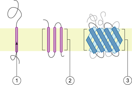
1) a single transmembrane α-helix (bitopic membrane protein).
2) a polytopic transmembrane α-helical protein.
3) a polytopic transmembrane β-sheet protein.
The membrane is represented in light green.