Magnification
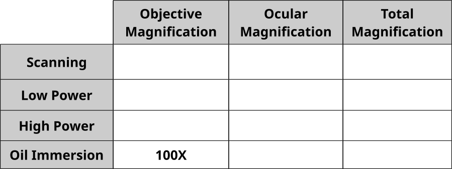
Fill in the following table. You can review magnification here.
Magnificationtotal = Magnificationobjective X Magnificationocular
Field of View Calculation
Follow the directions below. You can review field of view here.
- Examine a ruler under scanning magnification.
- Measure the diameter in millimeters (mm).
- Diameter = ______________
- Radius = _____________
- Calculate the field of view at this magnification = ______________
- Examine a ruler under low magnification (10x).
- Measure the diameter in millimeters (mm).
- Diameter = ______________
- Radius = _____________
- Calculate the field of view at this magnification = ______________
- What is the relationship in the between the magnification and field of view?
- What is the proportion of change in field of view when doubling the magnification?
The Letter e
- Follow this link: https://www.ncbionetwork.org/iet/microscope/
- Click on “Explore” → Click the sample box “?” → Click “Sample Slides”
- Click “Letter E”
- The slide is oriented so the “e” is right side up.
- If the image is blurry, use the focus sliders to make the image clear.
- What do you observe about the image under the microscope?
- Switch between scanning, low power and high power.
- Draw the “e” at scanning, low and high magnification.

Depth of Field
Follow the instructions to explore depth of field. You can review depth of field here.
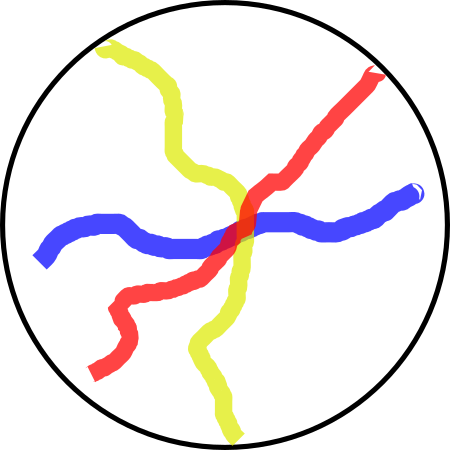
- Examine the slide of colored threads under scanning power so the cross-point of the threads is at the center of the field (see image below).
- Raise the magnification to the low power objective.
- What do we notice about the threads and the focus?
- How can we explain this observation with respect to the threads?
- Close the diaphragm so allow a pinpoint of light through the slide. What effect does this have on the image?
Examining Cells
- Choose a prepared slide of a Protist (Euglena, Amoeba, Paramecium).
- View the slide under 5x, 10x, and 40x magnification.
- How does the image change as you change magnification?
- Prepare a wet mount of a drop of pond water and place a cover slip over the drop.
- View the slide under 5x, 10x, and 40x magnification.
- Do you see anything moving?
- Prepare a slide of your own cheek cells.
- Swab the inside of your cheek.
- Roll the swab across a slide.
- Drop some methylene blue onto the slide.
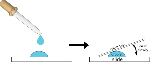
- Place a coverslip over the drop (see image below).
- View your cheek cells under 5x, 10x, and 40x magnification.
- Document your observations by drawing the cells and by using your phone to snap an image.