Table of Contents
Chromosomal Features
Chromosomes in interphase of the cell cycle are not visible individually. In preparation for nuclear division (mitosis or meiosis), they begin to organize tighter and condense in preparation for movement to subsequent daughter nuclei. The animation below illustrates the process of histone packaging and the molecular visualization of DNA replication. Histones are proteins that aid in packaging of the chromosomes into organized coils that give rise to the recognizable chromosomes during metaphase. This complex of DNA and proteins is called chromatin.

Humans have 23 pairs of chromosomes for a total of 46 chromosomes. The first 22 pairs of chromosomes are labeled 1 through 22 and are called autosomes. The last pair of chromosomes are the sex chromosomes. Individuals with two X sex chromosomes (XX) are female; those with one X and one Y (XY) are male.
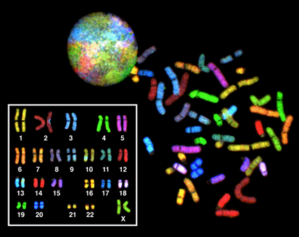
Karyotypes
Large-scale genomic rearrangements result in genetic abnormalities. Biologists utilize a technique called a chromosome spread followed by a karyotype or karyogram. To make a chromosome spread, one blocks the progression of mitosis at metaphase where chromosomes are condensed into the structures we are familiar. A karyotype analysis is an arrangement of the chromosome spread into the homologous pairs of chromosomes. The homologous pairs are arranged by size (largest to smallest). The sex chromosomes are placed last in the karyogram.