Table of Contents
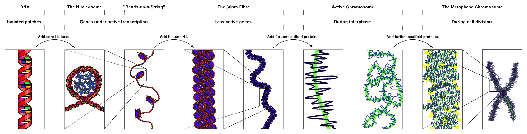
DNA condensing stages

Chromosomes are made of double stranded DNA molecules wound about histones and condensed into the familiar X-shape. Under regular functioning, these chromosomes are decondensed into filaments of chromatin in the nucleus and not recognizable.

Visualization of the chromosomes was done with Fluorescence in-situ Hybridization technique (top figure).
Chromosomes can only be seen when the cell is going to divide.
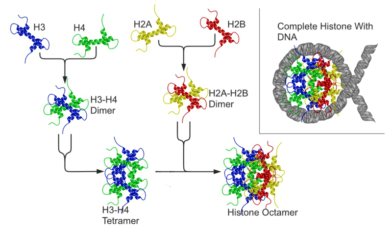
Histones
Chromosomes in Interphase are not visible individually and are loosely packaged within the nucleus. In preparation for nuclear division (mitosis or meiosis), they begin to organize tighter and condense in preparation for movement to subsequent daughter nuclei. The animation below illustrates the process of histone packaging and the molecular visualization of DNA replication. Histones are proteins that aid in packaging of the chromosomes into organized coils that give rise to the recognizable chromosomes during metaphase.
Nucleosome
DNA is negatively charged due to the Phosphate backbone. Histones are positively charged proteins that associate with eukaryotic DNA to spool and compact the DNA. The form into basic units called nucleosomes. The basic nucleosome core is composed of and octamer of histone proteins (H2A, H2B, H3 and H4)