Table of Contents
Cell size
Watch this video about what governs the size of a cell. How big can a single cell be?
Range of structure sizes and how we can see them. For example, mitochondrion and bacteria are about the same size (1 micrometer length) and can be seen using electron and light microscope.

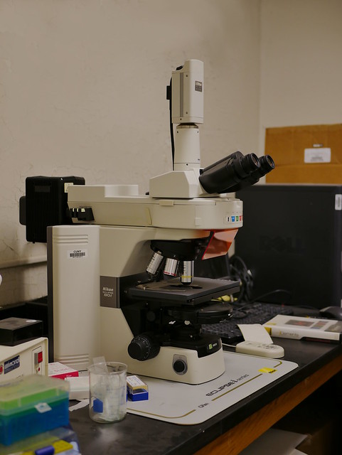
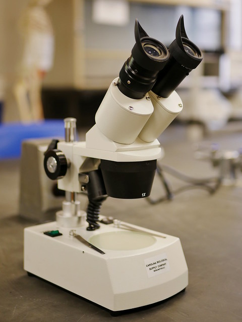
Modern compound microscope

Unlike van Leeuwenhoek’s single lens microscope, we now combine the magnifying power of multiple lenses in what is called a compound microscope.
Electron microscopy

An electron microscope is a microscope that uses a beam of accelerated electrons as a source of illumination.
Scanning and transmission electron microscope has magnifications of up to about 10,000,000x whereas most light microscopes have a limit of magnifications below 2000x
Scanning electron microscope (SEM) shows pictures of structures in 3D while transmission electron microscope (TEM) gives pictures of structures in 2D.

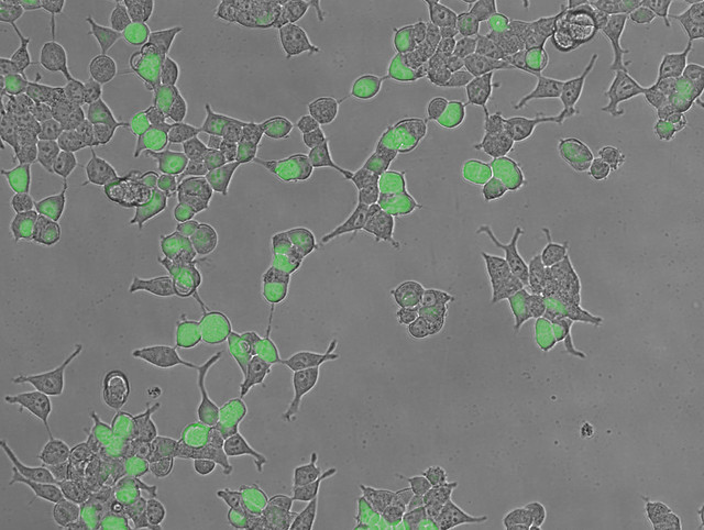
Fluorescent microscopy

- Antibodies developed against a specific protein
- Fluorescent dye molecule attached to antibody molecules
- Specimen exposed to fluorescent antibodies
- Ultra-violet light (black light) passed through specimen
- Fluorescent dye glows in color where antigen is located
- Emitted light is focused by glass lenses onto human retina or camera
- Allows mapping distribution of a specific protein in cell