Case 1- Periapical Cemental Dysplasia
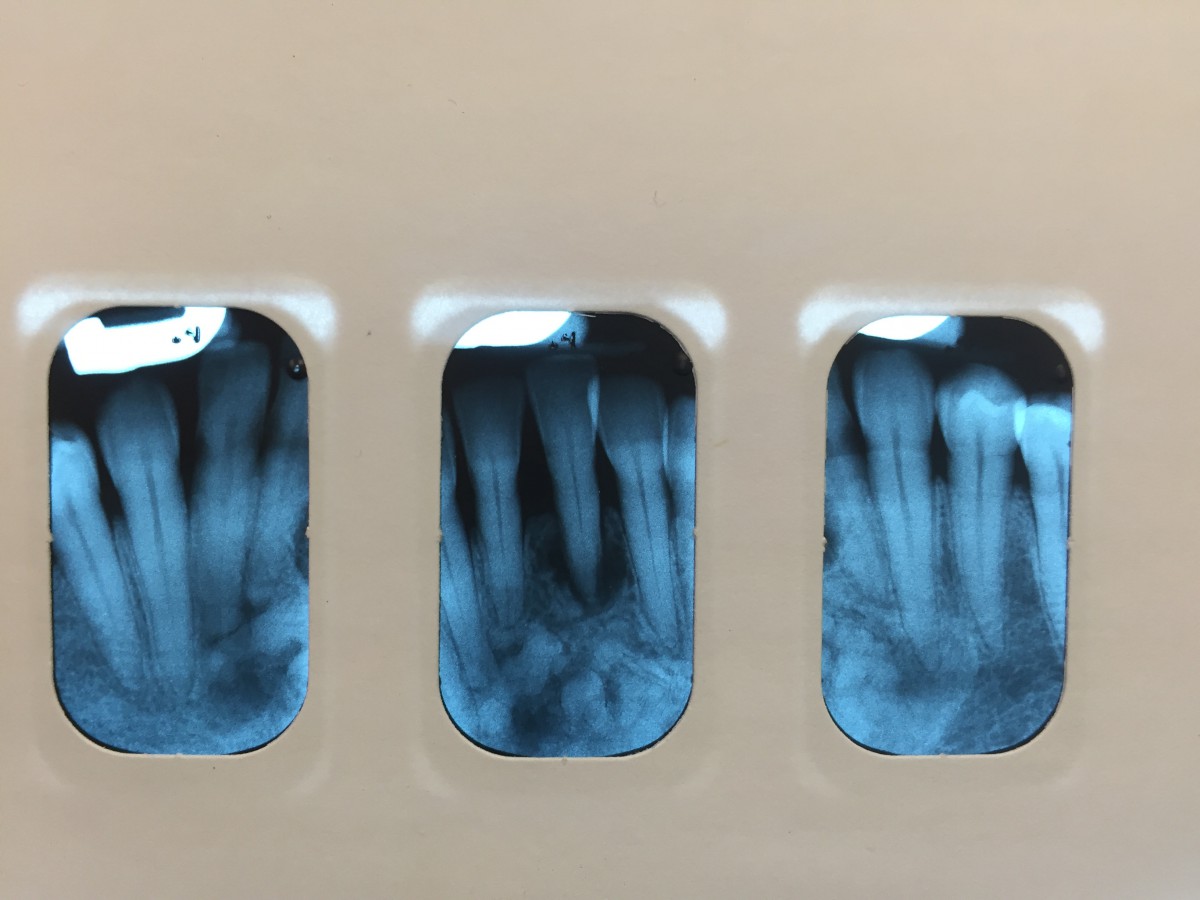
This patient was a middle aged African-American female. Dr. Brown was able to diagnose, from radiographs, that the patient presented with periapical cemental dysplasia. The patient was aware of this from previous visits to the dental clinic at City Tech. However, tooth #24 is discolored with more than 3mm of bone loss around it. The patient was told that it should be extracted and the only way to replace the missing tooth would be to get a flipper. This patient would not qualify for implants because of the bone abnormality.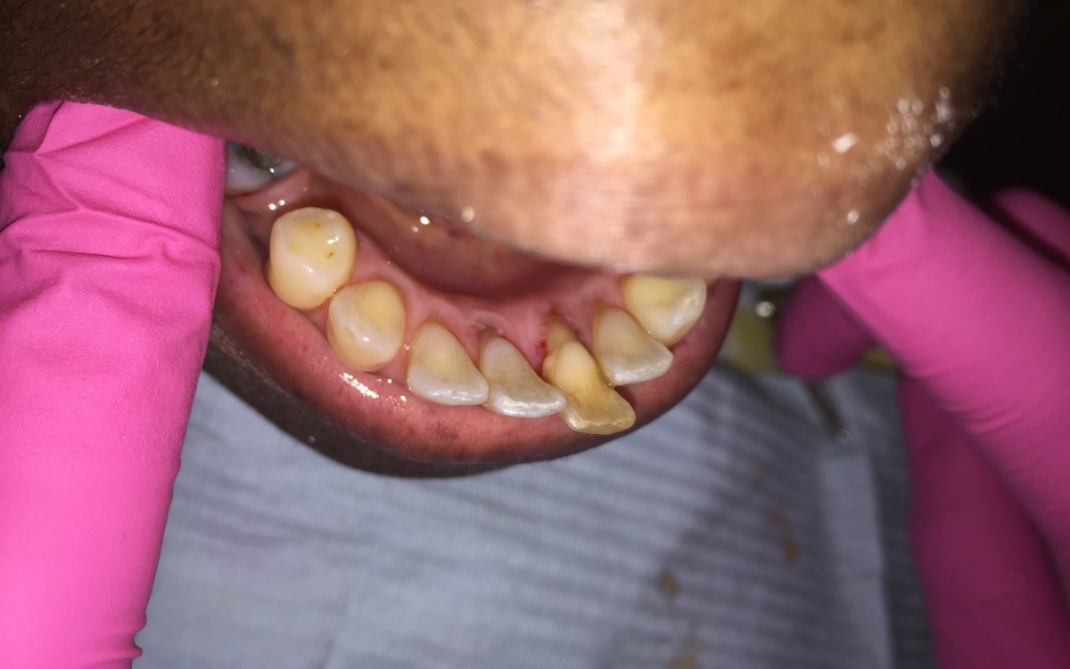

Case 2- A Regular Clinic Day with a Heavy Patient
These are some pictures from a typical dental cleaning on a heavy patient who is also a cigarette smoker. Note the staining and supragingival calculus present in the first picture. After a cleaning at the dental clinic, patients will leave with a white clean smile free of calculus and plaque.

Case 3- Pyogenic Granuloma
My very first patient of fourth semester presented with a pyogenic granuloma. Both Dr. Brown and Dr. Bowers had confirmed this diagnosis, although typically a biopsy is needed. A pyogenic granuloma is a small, round, bloody-red in color, growth that can occur intra-orally or extra-orally. In this case, the lesion presented intra-orally. They are very vascular and tend to occur in pregnant woman, although my patient was not known to be pregnant at the time. These lesions are benign and can be removed through surgical excision. This patient was not even aware of the lesion and said that it did not cause any pain. I debrided the area and gave her a referral for evaluation by an oral surgeon.


