
This project/paper was developed and researched in my oral pathology class during my 3rd dental hygiene semester. The paper discusses the etiology of a mucocele, how it is diagnosed, treated and it’s prognosis. This research paper helped develop my clinical understanding of certain pathological findings and how to recommend proper care of this case.
Ranula
By Naila Legros
Overview
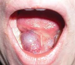
Ranula is a mucocele, or a mucus cyst involving a sublingual gland through the mylohyoid muscle dehiscence. Ranula is classified as a salivary gland disorder where swelling is observed on the neck and/or the floor of the mouth and occurs as a result of trauma or obstruction to the salivary gland, excretory duct and spillage of mucin into the surrounding soft tissues. It typically presents as a translucent blue, large palpable lesion, where resultant elevation of the tongue may interfere with swallowing. This cyst can fluctuate in size, making it difficult to detect and may be asymptomatic. Ranula’s have often been confused for a midline dermoid cyst, but due to its distinct location, it can be identified.
Etiology
The minor salivary glands are the most frequently injured glands of the lower lip. The etiology of ranula is due to a trauma where tissue gets caught during mastication, having a habit of lip biting or tongue thrusting. The trauma results in the detachment of the excretory duct and a leakage of mucus into the nearby tissue of the minor salivary gland.
Trauma during birth may affect the oral cavity as well, and cause some congenital mucoceles to newborns. Contributing factors include the baby sucking their fingers in utero as well as the baby passing through the birth canal.
Clinical Presentation
In earlier times, the name ranula derived from a latin word, “rana,” meaning frog. Ranula has been described as a little frog, due to the mucous cyst’s resemblance of the bulging underbelly of a frog.
In a case study, where a physical examination revealed a mandibular torus as well as a soft, compressible, mobile lesion. The mass did not move with the protrusion of the tongue. The lesion was described as a benign neck lesion with many differential diagnoses.
Demographic
In a recent ten year study from 2006-2016 had demographics of 3:1 female to male ratio. The age ranged between 3rd and 4TH decades of life, where the mean age of this study was 28 years old. 50% of the patients were predominantly of Polynesian descent. However, in a later study, there were no sex or age predilection.
Biopsy / Histology / Radiographs
Ranula’s can be diagnosed through a histological examination and through radiographic technique. Using a radiographic technique to diagnose ranula, one must take a radiograph that exposes the sublingual space.
In a histological examination, ranula appears and consists of a central cystic space containing mucin with a pseudocyst wall with loose vascularized connective tissue. An important feature when diagnosing ranula is the absence of epithelial tissue in the walls of ranula. A biopsy of the cystic walls is always recommended to rule out the possibility of a squamous cell carcinoma.
Differential Diagnosis
Mucoceles such as ranula beneath the tongue can often be confused for other pathological findings. As mentioned before, a squamous cell carcinoma is a typical differential diagnosis, as well as hemangioma, dermoid cyst, lymphangioma, benign/ malignant salivary gland neoplasm, lipoma, venous lake or an abscess.
Treatment
To treat any mucoceles or ranula, a surgical excision is the most commonly known treatment. Due to the lesion usually being rather small, a complete excision with the associated salivary gland is preferred. It is uncommon for the lesion to recur once the affected salivary gland has been removed.
An alternative treatment would be a corticosteroid injection along with a cryogenic therapy to the area.
Prognosis
As stated before, Ranula tends to be painless or asymptomatic with little or no associated mortality. Varying on location and size, some ranula may interfere with mastication, swallowing, speech and airway obstruction. If a thoracic ranula is present, it may alter and potentially compromise functions in respiration and may be life-threatening. If a complete excision of the ranula is reached, there should not be a recurrence.
In the case of a minor salivary gland ranula, if the gland is removed or transected, there is an increased risk of recurrence. In pediatric research, recurrence rates range from 5-10% post operation. Ranula lesions usually appear 6-8 weeks post surgery, however, a full recurrence is found within 12 months. With sufficient surgical excision, these mucus retaining cysts are not likely to recur.
Professional Relevance
Although dental hygienist’s main concern is preventative dental care, knowledge of oral pathology such as ranula is relevant to a dental hygienist. During extraoral and intraoral examinations, noting the presence of a lesion like ranula is necessary to help the patient retrieve proper treatment from either a general dentist or a specialist such as an oral surgeon.
Citations
- Burkhart, Nancy. “Ranula: A Review of Literature.” RDHMag.com, 1 Aug. 2009, rdhmag.com/articles/print/volume-29/issue-8/columns/oral-exams/ranula.html.
- Cheng, Adam. “What Is In Your Mouth.” Case 1: What Is in Your Mouth, 3 Nov. 2016, ncbi.nlm.nih.gov/pmc/articles/PMC2435329/
- Lomas, Jonathan, et al. “Surgical Management of Plunging Ranulas: a 10‐Year Case Series in South East Queensland.” The Canadian Journal of Chemical Engineering, Wiley-Blackwell, 21 Dec. 2017, onlinelibrary.wiley.com/doi/full/10.1111/ans.14356
- Verma, Gaurav. “Ranula: A Review of Literature.” Https://Pdfs.semanticscholar.org/Cdbf/3b318580cc4be61a440e14419f786b82e923.Pdf, 1 Oct. 2017

