Patient profile
Mrs. A is a 45 y.o. Indian woman presented for a dental cleaning. Married, non-smoker, no allergies reported, and no alcohol consumer. The patient states she uses a medium manual toothbrush 2x/day, Sensodyne dentifrice, flosses 2x/week, uses mouthwash rinses 2x/week.
Chief complaint: “I am due for a dental cleaning, and I need a deep cleaning as suggested by the previous hygienist”
Vitals: BP: 119/79, P:77 corresponding to normal.
Med/dental Hx: Reports no hospitalizations or medications for any medical conditions in the last 5 years. The patient states she has Vitiligo, and it manifests as white pouches on the skin. Reports she is taking OTC Vitamin D 2000IU/day. Her last physical exam was 6 months ago. Her last dental checkup was two years ago had 4BWs radiographic images taken, her last dental cleaning was a year ago.
ASA 2 ( due to Vitiligo condition)
Clinical Findings
Extraoral/Intraoral findings: Pale white patches of mission melanin on the lips. Bilateral linea alba. Palatal torus. Moderate redness on tonsils. Occlusion: molar relation Class I bilateral. Overjet- 3mm. Overbite- 10%. Generalized attrition noted. Gingival statement: generalized moderate inflamed red color of the gingiva, fits enlarged around the teeth, interdental space is filled with pyramidal shape papilla, localized blunted papilla on mandibular incisors, rolled gingival margin, soft-friable gingiva consistency, and smooth-shiny texture. Calculus: Localized supragingival calculus and light brown stains on mandibular incisors and heavy subgingival interproximal calculus.
The findings from the radiographic images were discussed with the patient and referrals were given to see a dentist.

Dental Charting:
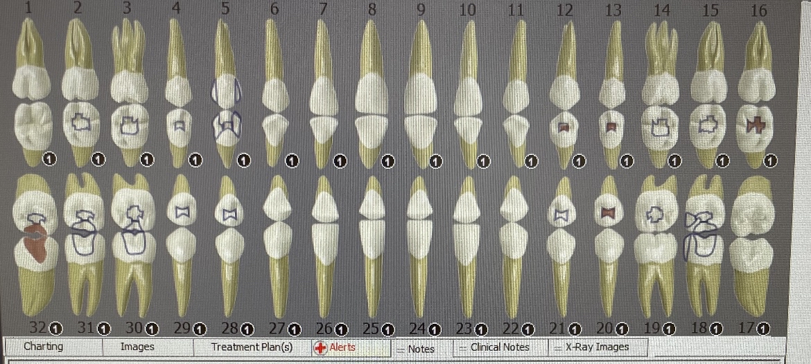
- Composite restorations on #2-O, #3-O, #4-O, #5-MOD, #12-O,#13-O, #14-O, #15-O, #16-O, #18-MO, #19-O, #20-O, #21-O, #28-O, #29-O, #30-OB, #31-OB, #32-O
- Caries lesions on #12-O, #13-O, #16-O, #20-O, #32-B
Periodontal Charting:

- Generalized moderate bleeding on probing
- Proping depth ranges between 2-3 mm on anterior teeth and 4-6 mm on posterior teeth.
- General gingival recession of 1-3mm
Periodontal Diagnosis: Periodontitis Stage II/Grade B (with radiographic evidence). Caries risk– High due to active caries lesions. Case value–High (due to heavy interproximal calculus).
Dental Hygiene Care Plan
Visit 1: PI/OHI- introduce the spool floss technique. Apply Oraqix (2.5%/2.5% lidocaine/prilocaine) for pain management. Ultrasonic and hand scale Q1. Give referral to see a dentist for #12-O, #13-O, #16-O, M, #20-O, and #32-B.
Visit 2: Take FMS radiographic images. PI/OHI- introduce the powered toothbrush technique. Apply Oraqix (2.5%/2.5% lidocaine/prilocaine) for pain management. Ultrasonic and hand scale Q4.
Visit 3: PI/OHI-proxy brushes for anterior incisors interproximal spaces. Apply Oraqix (2.5%/2.5% lidocaine/prilocaine) for pain management. Ultrasonic and hand scale Q2 and Q3. Apply 5% varnish fluoride.
Implementation
1st visit summary: All intake data was collected, and the Tx plan was signed by the patient. PI/OHI- introduced spool floss technique. Applied Oraqix (2.5%/2.5% lidocaine/prilocaine) for pain management. Ultrasonic and hand-scaled Q1. The referral was given to see a dentist for #12-O, #13-O, #16-O, M, #20-O, and #32-B caries lesions. Residual calculus was noted on #2, 3, 4, 5-D, #5-M, and #7-M.
2nd visit summary: FMS radiographic images were taken and discussed the finding with the patient. Re-evaluated scaled Q1 on the last visit. The patient had some improvement in tissue response on scaled Q1, a decrease in inflammation of the gingiva was noted, pale pink color gingiva, fits snugly around the teeth, pyramidal shape papilla, still enlarged around teeth #1 and #2 . Mild bleeding on exploring. PI/OHI- introduced powered toothbrush technique. Applied Oraqix (2.5%/2.5% lidocaine/prilocaine) for pain management. Ultrasonic and hand-scaled residual calculus on #2, 3, 4, 5-D, #5-M, #7-M, and Q4. Residual calculus was noted on #27-D and #29-D.
3rd visit summary: Re-evaluated scaled Q4 on last visit. Gingival tissue response: was noted significant improvement, pink gingiva color, fits snugly around the teeth, rolled gingival margin, pyramidal shape papillae, and no bleeding on exploring, localized soft gingiva consistency around #30-32. Supragingival calculus was noted on #25-27. PI/OHI-re-inforced powered toothbrush technique and introduced proxy brushes/ patient was able to use it properly. Applied Oraqix (2.5%/2.5% lidocaine/prilocaine) for pain management. Ultrasonic and hand-scaled residual calculus on #27-D, #29-D, Q2, and Q3 to completion. Applied 5% varnish fluoride with post-op instructions.


Continued Care Recommendation: 3 months of re-care was recommended to the patient due to the diagnosis of periodontitis, heavy subgingival calculus, and supragingival calculus accumulation.

