Initial Visit
- 28 year old African American female.
- Chief complaint: In need of a dental cleaning.
- Vitals: 116/85 P: 81, corresponding with Stage 1 hypertension
- Patient reports being allergic to peanuts
- ASA II
Assessments
- EO findings: Patient presents with swelling on the right aspect of her thyroid gland.
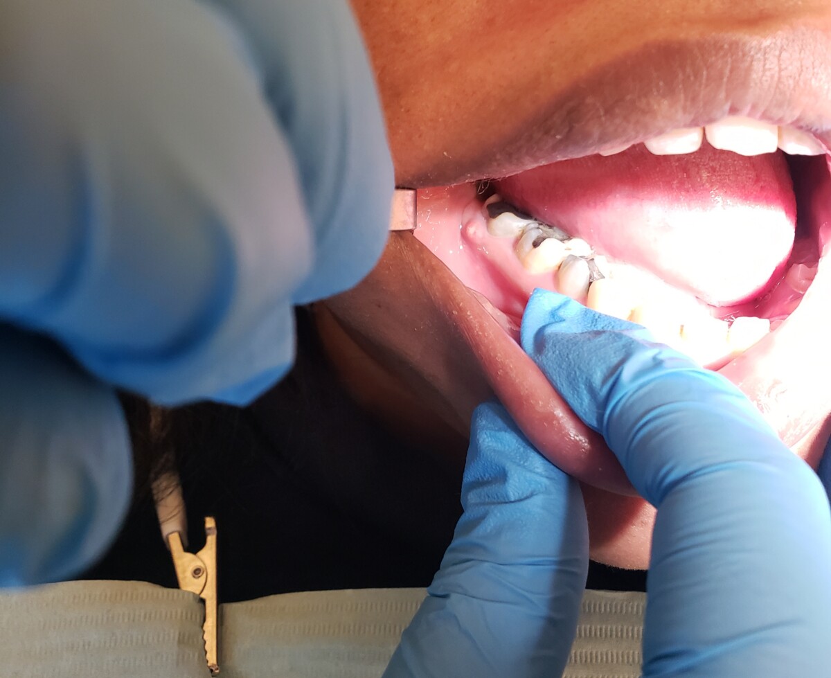
- IO findings: the patient presented with a fistula on the buccal aspect and apical to #30. It is approximately 5mm in size, attached with a regular border, and is red in appearance with pus visible at its center.
- Bilateral mandibular tori and linea alba
- There is a short attachment of the lingual and labial frenum.
- Overjet: 5mm, Overbite: 20%, bilateral class I occlusion.
Dental Charting
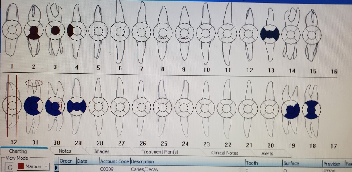
- #16 and #17 are clinically missing. Patient reports that they were not extracted. After review of her radiographs, it was observed that #16 was congenitally missing and #17 was unerupted with a cyst formed around it.
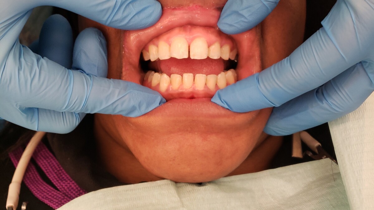
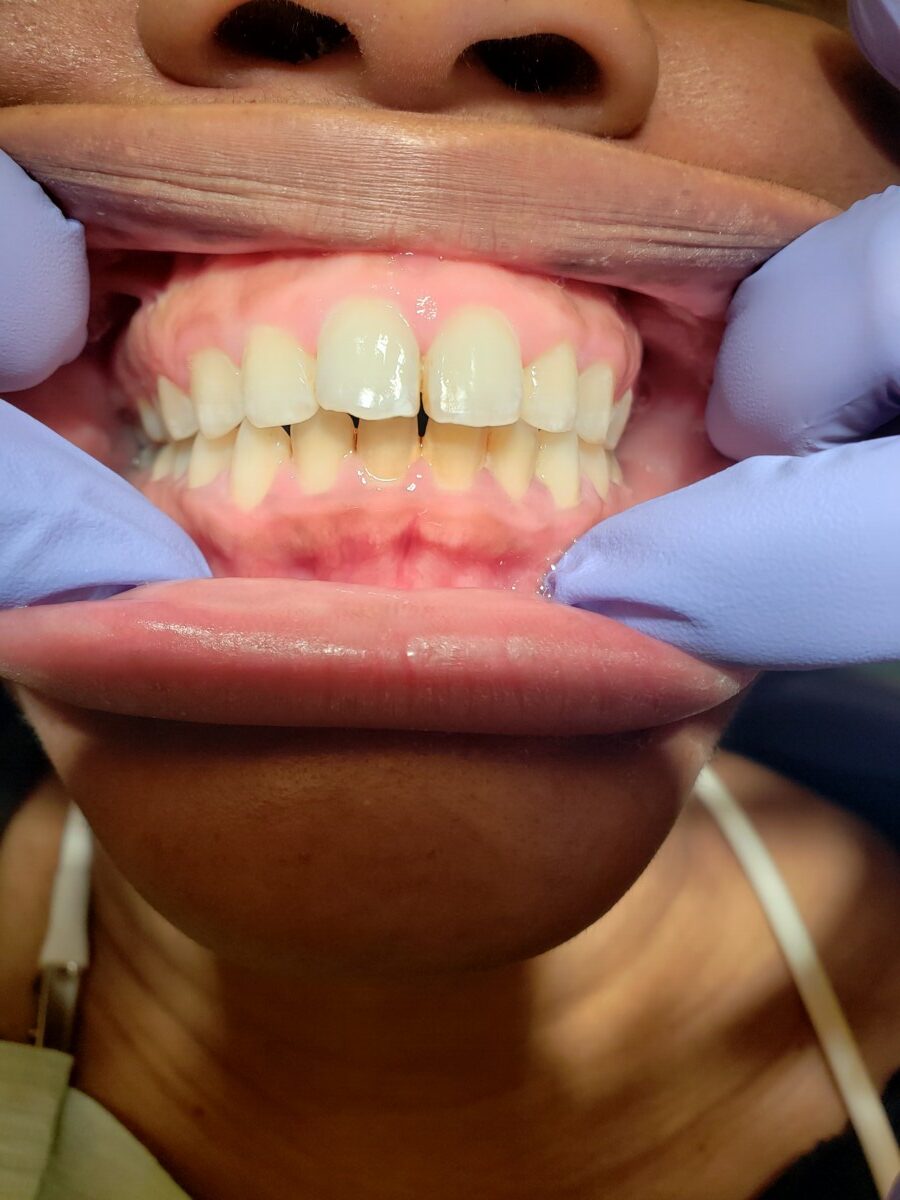
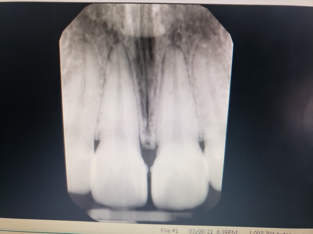
- Fractures observed on the incisal edges of 8 and 9, as well as on #30M.
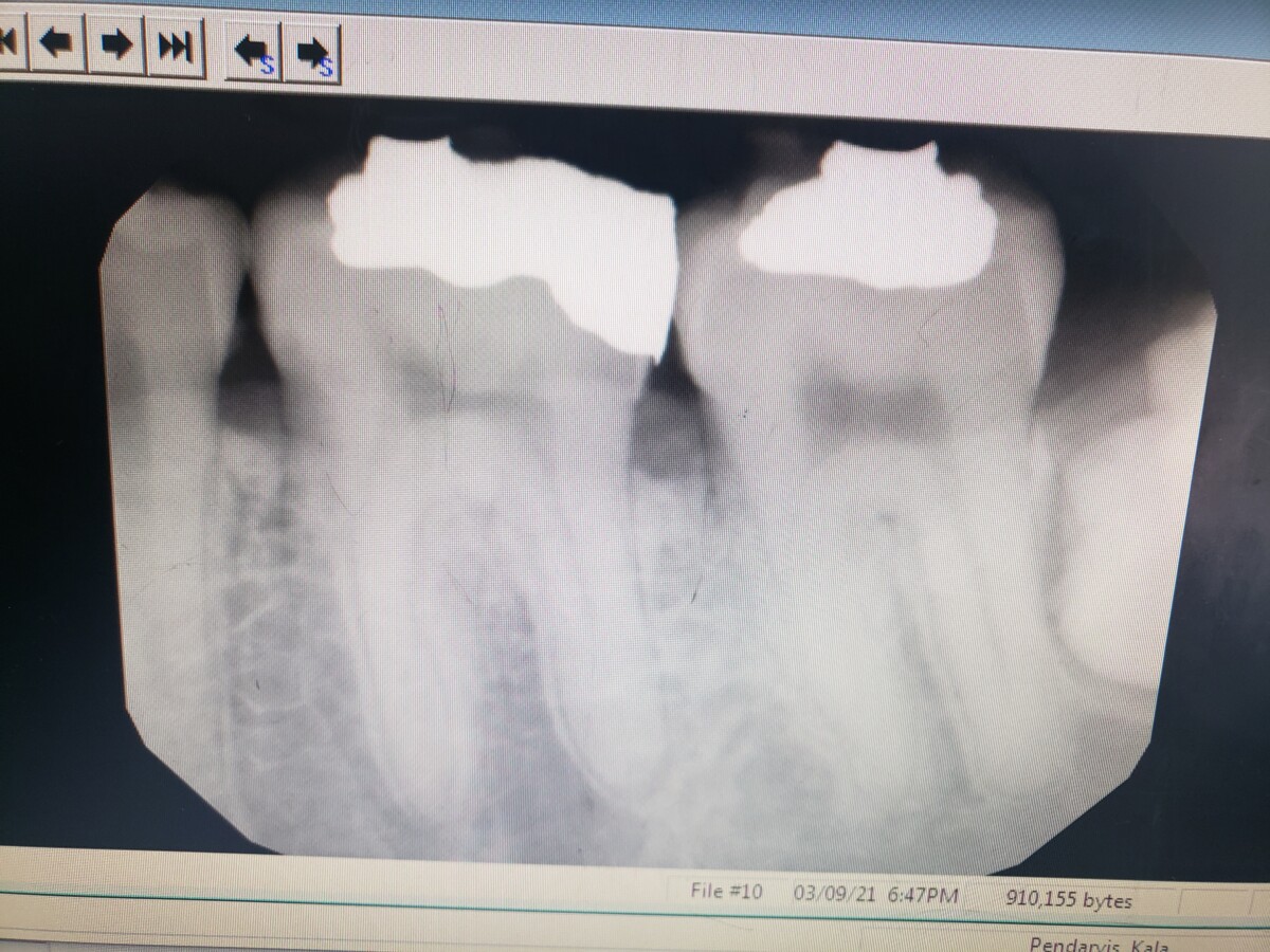
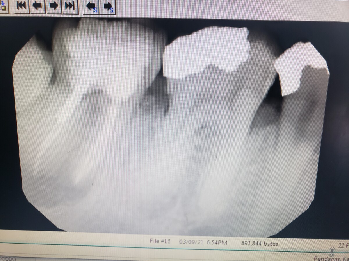
- Amalgam restorations: #13-MOD, #18-OBL, #19-ODB, #29-MOD, #30-ODB, #31-ODBL
- Suspicious carious lesions: 2OL, 3O, and 4D
- #32 is partially erupted.
Periodontal Charting
- Localized, moderate BOP.
- Generalized probing depth of 1 to 4mm
- Localized areas on the posterior teeth with probing depths up to 7mm.
- Note that teeth #s 30 and 31 were not probed in order to avoid the fistula that was observed during IO exam.
- No recession observed, possibly due to the erythematous nature of the gingiva.
Gingival Description
The patient’s gingiva presents as pink with some stippling, however, there is mild, marginal inflammation present. The interdental papillae are dark pink, erythematous, and lack resiliency. Patient also states that she is feeling and intolerable level of pain during probing and exploring.
Radiographs
- FMS exposed at 7mA, 70 kVp.
- Generalized horizontal bone loos observed, approximately 15%. This corresponds with Stage 1 periodontitis.
- Periapical pathology seen at the apices of #s 19, 30, and 31.
- Suspicious lesions observed on 3-M, 4-D, 13-D. #s 19, 30, 31 also show signs of recurrent decay.
- A cyst has formed around #17 which is unerupted.
Diagnosis
- Patient has been determined to have Stage 1 periodontitis due to her level of bone loss. Grade B because the destruction of the periodontium proportional to the amount of biofilm deposits.
- High caries risk with active lesions and recurrent decay.
- Generalized, heavy subgingival calculus deposits and localized, heavy supragingival calculus along the mandibular anterior. Heavy case value.
Treatment Plan
- Visit 1: Complete assessments and expose FMS.
- Visit 2: Scale quadrants 1 and 4 using local anesthesia. Avoid teeth numbers 30 and 31 due to fistula and PAPs observed on radiographs.
- Visit 3: Scale quadrants 2 and 3 using local anesthesia. Avoid teeth #s 18 and 19 due to PAPs observed on radiographs. Engine polish with medium grit prophy paste. Apply 5% sodium fluoride varnish.
Treatment Plan Implementation
- Visit 1: Assessments completed. FMS exposed. PI: 1.8. OHI: Introduced Bass method of tooth brushing. Referral was giving to patient for evaluation of the swelling to the right of her thyroid, her high BP readings, the suspicious lesions observed in her radiographs, and the fistula adjacent to #31.
- Visit 2: PI: 1.2. Introduced pt to proper flossing technique using floss picks. Hand scaled quadrants 1 and 4. Used 1 carpule of 2% Lidocaine with 1:100,000 epinephrine via ASA, MSA, and PSA infiltration. Negative aspiration for all 3. Vitals before administration: 126/77 P: 75. Vitals after administration: 130/88 P: 79. 20% benzocaine was applied to injection area before administration. Patient tolerated treatment well.
- Visit 3: Scaled quadrants 2 & 3 using 1 carpule of 1:100,000 epinephrine via MSA, PSA, and mental infiltration. Negative aspiration for all 3. Vitals before: 125/78 P: 78. Vitals after: 128/81 P: 82. Engine polished using medium prophy paste. 5% sodium fluoride treatment applied. Aftercare instructions given,
- Next visit in 3 months.