Initial visit
- 31yo African American male
- Chief complain: Gingival bleeding and in need of a cleaning
- Vitals: BP: 126/75 P:74
- Smoker for the past 10 years about 10 cigarettes/day
- ASA: II
Assessments
- EO: No significant findings
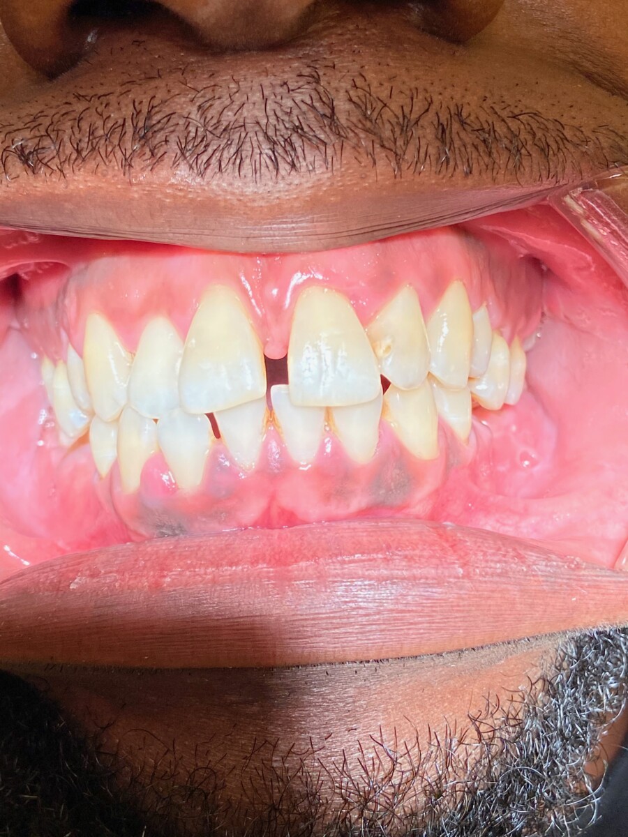
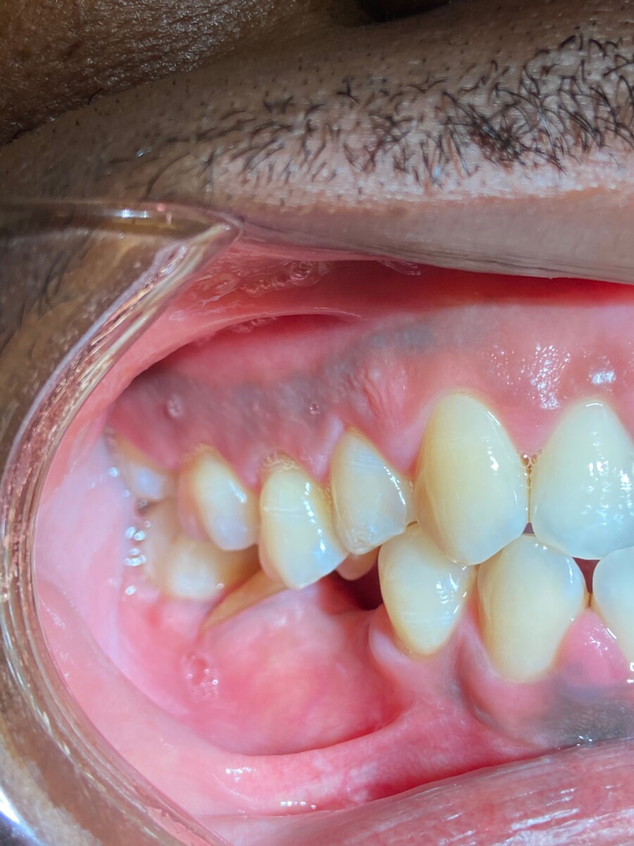
- IO: Melanin pigmentation of the gingiva, Palatal torus, Bilateral slight lines alba on the buccal mucosa, Oropharinx slightly inflamed.
- Class of occlusion: Bilateral Class I with #7 & # 26-edge to edge bite, Overjet: 4mm, Overbite: 10%
Dental charting
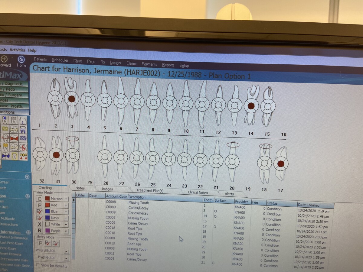
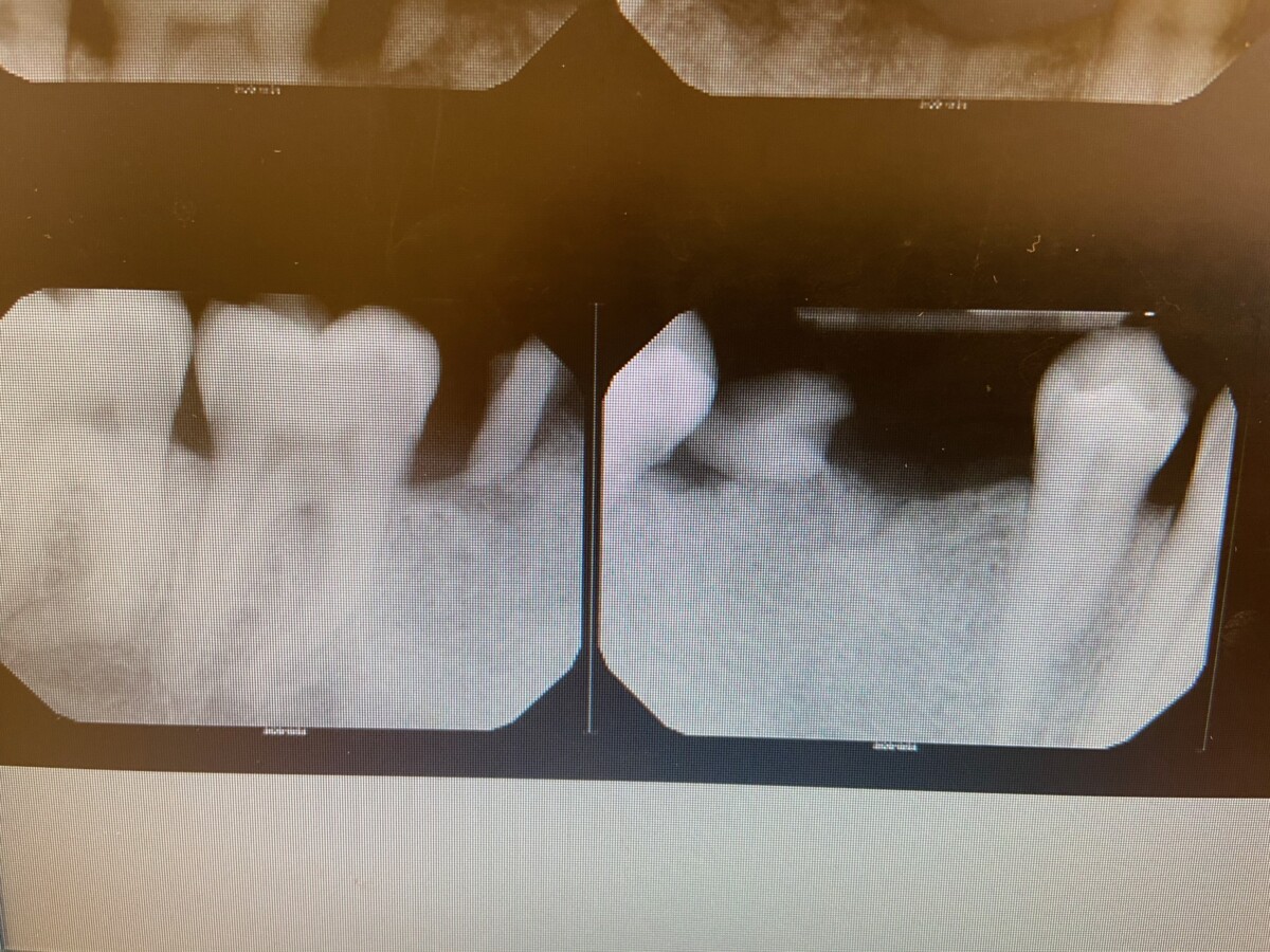
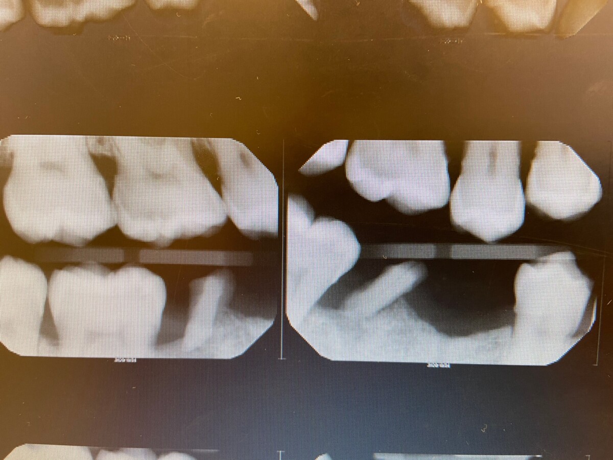
- Missing teeth #29-extracted due to big carious lesion
- Retained roots: #18, #19, #20 ,#30
- Patient present with no attrition
- Carious lesion present on #3, #14, #17 and #31.
- #1 & #16 are not clinicaly present.
Periodontal probing
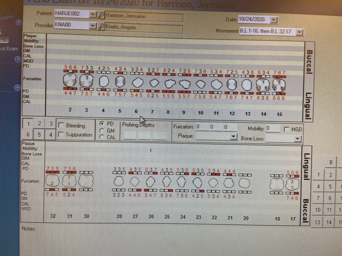
- Generalized heavy BOP
- Generalized probing depths of 1mm-8mm
- Note # 18, #19, #20, #30 were not probed due to being retained roots.
Gingival Assessment
- Generalized gingival inflammation, red erythematous in color with melanin pigmentation, generalized bulbous papillae red-blush in color with rolled gingival margins.
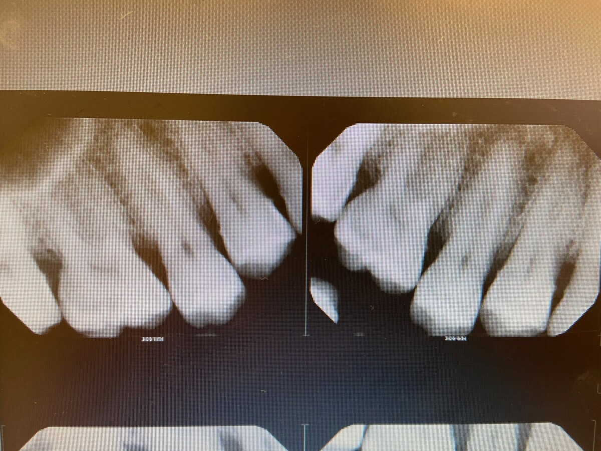
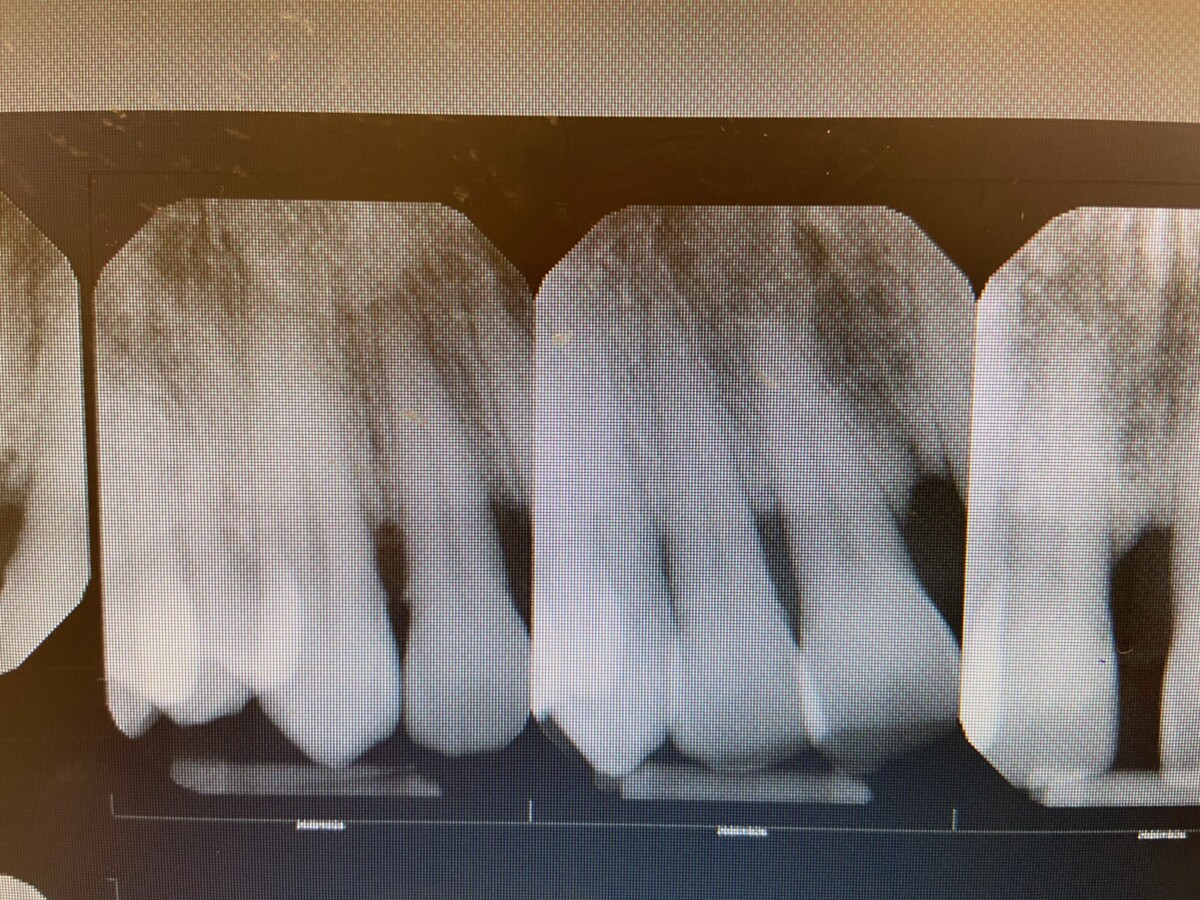
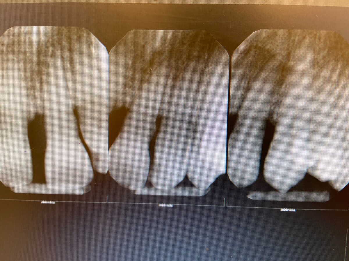
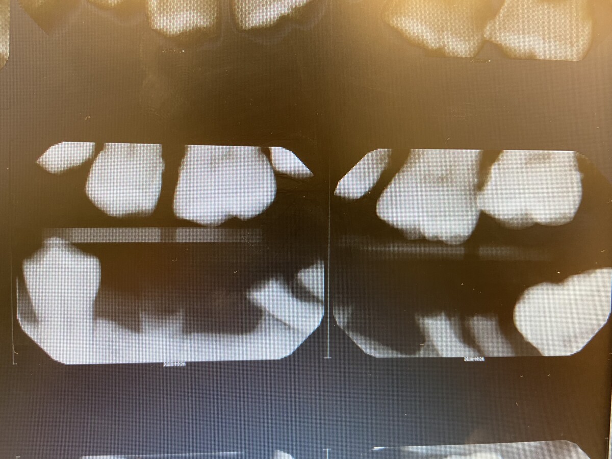
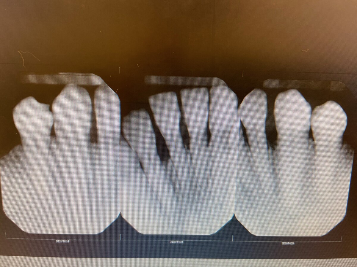
Radiographs
- FMS exposed at 7mA, 70kVp.
- Generalized horizontal bone loss was observed of about 30%. This corresponds with Stage 3 periodontitis.
- Suspicious lesions confirmed with radiographs
- Retained roots confirmed with radiographs
- Referral was given to patient for the extraction of the retained roots, evaluation of the suspicious lesions as well as possible implants for the missing teeth.


Diagnosis
- Patient has been diagnosed with Stage III periodontitis due to levels of bone loss. Grade B due to the level of periodontium destruction as well as patients smoking habits of 10 cigarettes/day.
- High risk caries due to active lesions present
- Generalized heavy sub and supra gingival calculus.
Treatment Plan
- Visit 1: Complete assessments, Expose FMS.
- Visit 2: OHI, Start scaling UR & LR using local anesthesia
- Visit 3: OHI, Start scaling UL & LL using local anesthesia, Engine polish, apply fluoride varnish
Treatment implementation
- Visit 1: Finished all assessments, exposed FMS. P.I.: 1- Patient was educated on the importance of flossing and was taught a proper flossing technique due to most of the biofilm was present at the gingival margin.
- Visit 2: P.I-1- Flossing method was reinforce and Listerine Total Care was recommended for remeneralization of the teeth an prevention of future carious lesions. Scaled UR & LR quadrant. Local anesthesia; IAN block was administered by the doctor on floor as well as PSA, MSA, ASA using 2 carpules of Lidocaine HCL 2% with 1:100k epi.
- Visit 3: P.I: 0.6- Patient stopped smoking, Toothbrushing technique was implemented- Modified Bass. Scaled UL & LL quadrant. Local anesthesia; IAN block was administered by the doctor on floor as well as PSA, MSA, ASA using 2 carpules of Lidocaine HCL 2% with 1:100k epi. Engine polished using medium grit prophy paste, applied Fluoride Varnish 5% NaF.
- 3 Months recare.

