Case Study #1: Irregular Eruption (L/I)
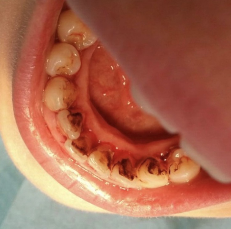
12 year-old caucasion male with a past history of GERD in which the parent reported he is presently taking Prilosec and the condition is well-managed. I proceeded with asking the parent if the child had any other symptoms of GERD such as frequent sore throat, dysphagia, chest pain, etc. in which the parent reported he did not have any symptoms associated with the condition. No allergies/recent hospitalizations or any other medical condition were reported. Although the condition was stable, consideration was later taken and the patient remained in a semi-reclined position during the entire treatment.
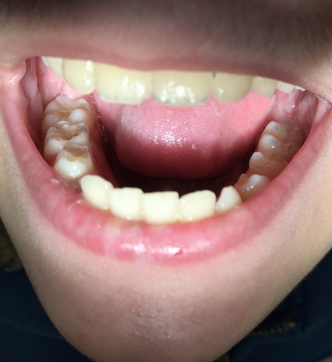
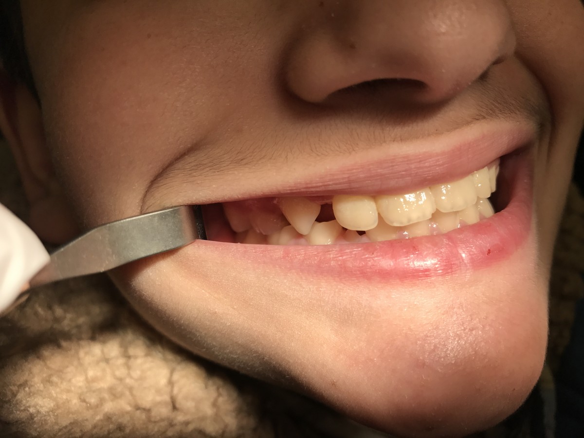
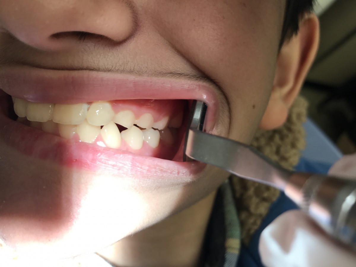
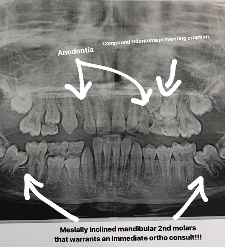
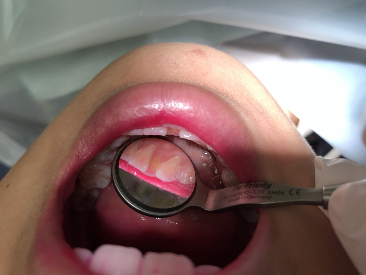
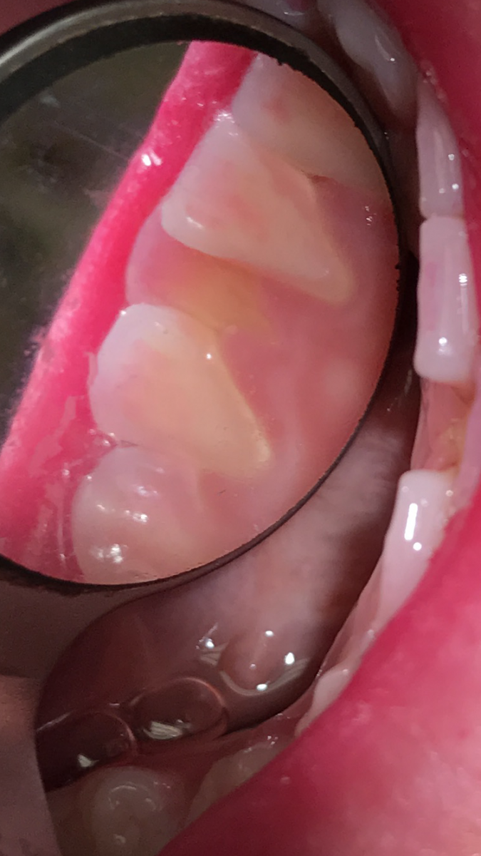
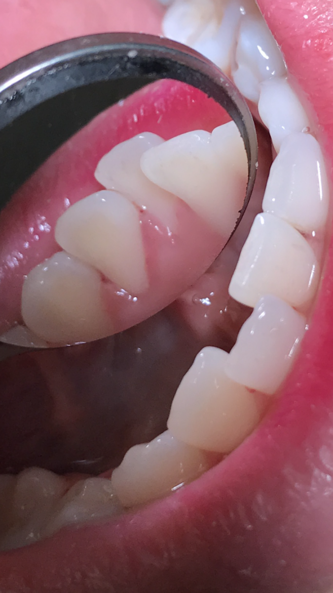
Treatment rendered: EO/IO was within normal limits although this was when I first noticed that the teeth (specifically the primary dentition) were eroded (since the parent did not mention the condition upon arrival). I then assessed the eruption of the permanent dentition. I exposed a PAN after clinical presentation showed some serious irregularities in his eruption pattern. His X-ray revealed anodontia of the permanent 1st premolars, a compound odontoma preventing the eruption of his permanent 2nd premolar & 1st molar, as well as unerupted mesially inclined mandibular 2nd molars that warranted an immediate orthodontic consultation. Clinical and radiographic findings were thoroughly discussed with the parent and I stressed the importance of seeking immediate dental care. I explained that if she didn’t seek proper care for the child, the mandibular 2nd molars would never erupt due to their position, #13 & #14 would not erupt either unless the odontoma was removed by an oral surgeon, the spaces due to the anodontia of the maxillary 1st premolars would result in the teeth mesially shifting and thus, occlusal function would be compromised. The worst part is that this patient had visited a dentist 1 month prior to walking into our clinic. The parent was told that the child had 4 cavities present meanwhile no caries were detected. Appropriate referrals were given for an oral surgeon (for the removal of the compound odontoma) and an orthodontic consult to allow the mandibular 2nd molars to erupt. Patient was also provided with a copy of the radiograph.
I then continued with my assessments and treatment plan. Localized moderate inflammation in the UR quadrant with generalized minimal inflammation and generalized minimal bleeding was present. A high plaque score was revealed, in which I then discussed, assessed & instructed proper brushing technique. Oral prophylaxis was then given using hand instruments, followed by polishing with fine paste & applying a 5% NaF.
In addition to the treatment provided, I implemented the management of tooth erosion. I informed the parent and patient that keeping hydrated as well as using a fluoridated rinse would help prevent further acidic damages to the enamel.
A follow up phone call to the parent was made to ensure that the child get appropriate care, and the parent had reported that she had visited an orthodontist and had been working on seeking an oral surgeon in order to get proper treatment.
The patient came back for a follow-up visit 2 months later for the placement of cotton-roll sealants on #14 & # 19. I also assessed his brushing technique in which I noticed improved drastically, and he reported to be brushing 2x a day where he admitted to occasionally forgetting to brush before going to bed. Previously detected localized moderate inflammation in the UR quadrant had reduced during this visit.
Case Study #2: Crowded Teeth (L/I)
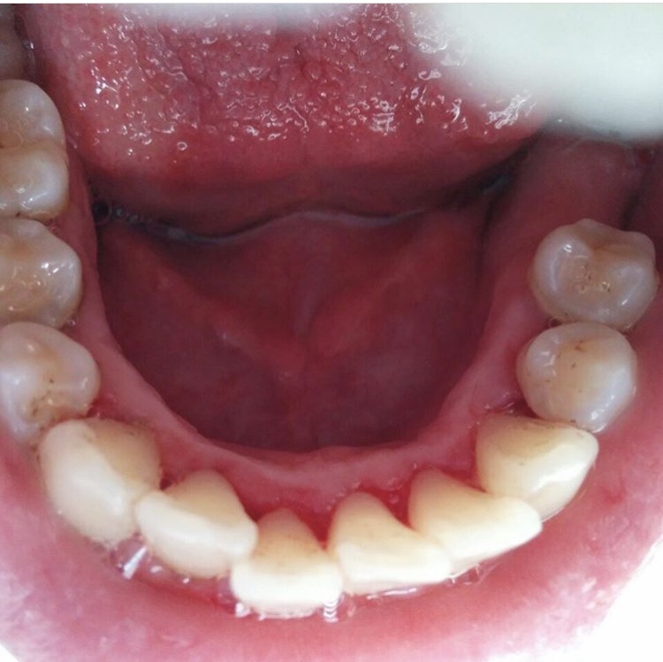
22 year-old asian female. BP 93/72 P77. Patient reported blood pressure was ‘normally’ within that range. Medical History was within normal limits. I found that the patient was taking vitamin C, D, and B12 as recommended by her MD due to vegetarian diet. Non-drinker and non-smoker. EO: TMJ clicking. Patient was aware but feels no pain. IO: No significant findings. Dental- suspicious areas of decay on the occlusal surfaces of #18 & #31 but due to the patient having recent X-rays taken, radiographs were not exposed at the clinic. Instead, I asked if her dentist reported any cavities present, in which she responded no. A referral was then given for the areas of decay found. Crowding was noted on #24-#26. Periodontal findings were generalized 1-3mm localized 4mm in sextants 4 & 6. Generalized minimal inflammation and generalized minimal bleeding present with localized moderate inflammation in mandibular sextants 4 & 6. Gingiva appeared generalized pink with localized red ‘band’ under molars of LR & LL quadrants. Generalized minimal subgingival calculus found with localized moderate supra and subgingival calculus on the mandibular anterior linguals were present due to crowding.
Plaque score was taken and revealed mainly interproximal plaque present. Recommended & instructed proper flossing technique and emphasized closer attention to area of crowding. Proper education on the implications of crowded teeth being a ‘plaque trap’ was used and shown to the patient with a handheld mirror.
Treatment rendered: V1- Assessments completed. Hand Scaled & Ultrasonic on all 4 quadrants. Polished with fine paste & applied 5% NaF. 6 month recare was given for this patient.
Patient was highly motivated with improving her oral hygiene routine. I explained that the area of crowding is where plaque and calculus build up most, and why it’s so important to clean that area in order to prevent the build up from occurring. I also explained that the red ‘band’ by the gingival margin around her molars in the LL & LR quadrants are areas where the gingiva is less healthy due to the plaque and bacteria present causing gingivitis.
Case Study #3 Staining (H/I)
28 year-old caucasian female. BP: 137/90 P68. High Blood Pressure fact sheet was given and explained to the patient. Patient stated that he recently went for a visit to his physician and BP was presently being monitored. No medications/ hospitalizations/or any other medical conditions present. Patient reported allergic to bee-stings with a mild reaction therefore he did not carry an EpiPen. Patient had a past history of smoking but reported that he had stopped smoking 3 months prior to the visit. EO: were within normal limits. IO: Nicotine stomatitis on the hard palate. Dental- multiple amalgams and composites present. #19 was missing. Generalized moderate subgingival and supragingival calculus present with generalized heavy lingual staining. Periodontal findings: Generalized minimal inflammation and generalized moderate bleeding present. Generalized 1-3mm with localized 4-5mm in sextant. #6 & #7 facials had 1-2mm of recession; patient may have been brushing too hard. Localized pale pink, bulbous & spongy tissue on mandibular anterior facials were noted. High plaque score was revealed: 1.5. Recommended & Instructed brushing technique using an electric toothbrush due to better plaque removal.
Treamtment provided: Assessments completed. Air polished all 4 quadrants. Hand scaled & ultrasonic all 4 quadrants. Applied 5% NaF. 3 month recall was given to heavy calculus and stain.
As mentioned, this patient had already quit smoking prior to the visit, so I congratulated the patient for this. Smoking cessation was then reinforced due to the heavy amounts of staining. I also felt that an electric toothbrush would be best to recommend due to the excessive plaque present and easier use.
Case Study #4: Arestin Patient (H/II)
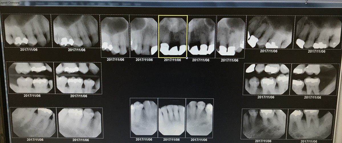
63 year-old african american male. BP 127/90. Medical History- Patient reported having a positive history of HSV II which was diagnosed in 1990, and has had no outbreaks since 1995 since stopping sexual intercourse. No medications were reported being taken. Patient is a non-drinker and non-smoker. ASA 2. EO/IO: reported no significant findings. Dental- multiple amalgams present. A 4-unit bridge was noted on #8-#11. All 4 1st premolars were extracted due to having orthodontic treatment in the past. Attrition was found on #7 & #23-#27. Periodontal findings reported localized fibrous tissue located in all posterior regions. In the residual areas, the gingiva was firm & resilient, non-stippled, and filled the interdental spaces. Gingiva appeared generalized brown due to melanin pigmentation. Generalized moderate inflammation and generalized minimal bleeding present. Generalized 2-4mm was noted with localized 3-9mm in the left posterior sextants. Generalized 1-3mm of recession on #14, #15. #18, #29 & #30; all reported to be asymptomatic. Localized advanced periodontitis was found in sextants 3 & 4. +2 mobility on #18. Arestin was later placed on this patient, however, in the areas where there is mobility and >7mm pocket depth in both sextants 3 & 4, patient was given a referral to see a periodontist. In sextant 1, #2 MB, & #3 MB & ML had 5mm of pocket depth where arestin was placed, after exposure of an FMS showed evidence extensive evidence of bone loss with severe bone loss around #18. It also revealed radiolucencies above the 4-unit bridge #8-#11 which I later added to the referral.
I then evaluated for calculus. Patient had generalized heavy subgingival calculus with localized moderate supragingival in the mandibular anterior lingual region. For homecare, I recommended the Modified Bass Method. In the subsequent visit, I recommended and instructed the use of thornton’s floss to thoroughly clean under the 4-unit bridge present.
Due to the heavy case value and deeper periodontal pockets, it took multiple visits to complete this patient. I used both hand instrumentation and an ultrasonic device to remove all the calculus present. I explained the implications of periodontal disease how to further prevent/manage the condition. After treatment was completed, I polished with fine paste and applied a 5% Fluoride Varnish. A 3 month recall appointment was recommended due to the patient’s oral condition.
During his second visit, I evaluated the gingiva for the response to treatment during the previous visit. The gingival tissue showed great positive changes. The tissue appeared significantly less fibrotic in the quadrants previously scaled and seemingly snug around the tooth unlike how it appeared before. It was also more resilient/firm and bleeding had slightly reduced. I then placed the arestin in the above mentioned sites. Post evaluation instructions were given.
The interval between the last dental visit and these visits was 18 years, therefore I strongly emphasized the importance of having a dental home. However, due to his financial situation, he had mentioned that he could not afford proper dental care. I therefore helped the patient find a low cost clinic and a follow up phone call confirmed that he had later found one and that he had an upcoming appointment scheduled. I also stressed the importance of maintaining proper oral home-care. He purchased all recommended aids & reported to have been using them.
