Case #1. Hypertension
A.K. 63 years old white male with a history of hypertension. Medium/ Generalized Type I and Localized Type II perio.
Initial visit. Last dental examination and radiographs were performed in 2015. Blood pressure was 162/80 (Patient is aware of high blood pressure. He stated that he visited cardiologist one day before hygiene appointment), pulse 67. ASA II. The patient takes Losartan 50mg daily for 3 years to control blood pressure. The major effect on dental treatment- orthostatic hypotension as patient stands up after treatment. To avoid this, the patient was advised to stand up slowly and was given a few minutes to sit in the dental chair in an upright position after the treatment was completed. The patient started to take Norvasc (Amlodipine) 5mg first day before the hygiene appointment, after visiting cardiologist. Amlodipine is a calcium channel blocker. Adverse effect on dental treatment- gingival hyperplasia. The patient didn’t have signs of hyperplasia during hygiene visit. The patient is allergic to penicillin. The patient smokes 1 1/2 pack of cigarettes per day for 30 years. Smoking cessation discussed.
Dentition: Occlusal class II, edge to edge bite. #15-MO decay and missing restoration, #24,25- fractured incisal edges, #21F- root caries. Referral for the evaluation of decay was given to the patient. Toothbrush abrasion present on #22,23,27.
Periodontal: The patient has pink, marginally rolled gingiva, non-resilient, with bulbous interdental papillae. Generalized probing depths 4mm, localized 5mm, BUP-moderate. Moderate calculus deposits with heavy staining contributed to smoking present. PI-0.8- considered fair.
Treatment management
All assessment data was collected, this included a review of medical history, BP reading, EO/IO exam, dental and periodontal charting, plaque index score and calculus detection, referral for the dental evaluation was provided. Upon careful review of my assessments, I developed an individualized treatment plan that would address his gingival condition. At the initial visit, I reviewed the proper flossing technique using waxed dental floss and floss threader. The patient was able to show the technique. The importance of smoking cessation and side effects of smoking was discussed several times during the appointment. During the appointment, I completed 4 quadrants scaling with ultrasonic and hand instruments. I polished entire dentition with a coarse paste to remove staining. 4 months recare was recommended to the patient.
Case #2. Arestin
M.A. 47 years old white male. Medium/ Type II perio.
Initial visit: The patient is in a good health condition. Vital signs were within normal limits, B/P 102/70, pulse was 59. No systemic conditions. No premedication needed. ASA I.
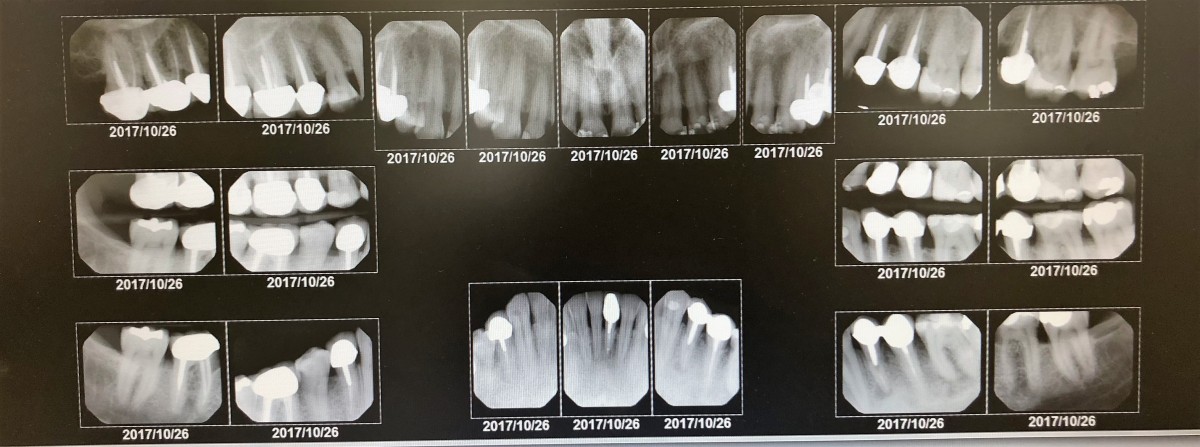
Oral Pathology: The patient did not have any visible oral pathology. The radiographs revealed unilocular radiolucency adjacent to the roots of teeth #21(2x4mm) & #24(2x1mm) presumably from failed root canal treatment. The referral for the dental evaluation was given.
Dentition: The patient has multiple restorations, all intact. Tooth #19 appears suspicious- dark under the composite. Radiographs revealed recurrent decay.
Periodontal: The gingiva is pink, marginally rolled, non-resilient, inflamed near the gingival margin. The patient was classified as periodontal Type II due to localized 5-7 mm pockets, generalized 3-4mm probing depths, grade 2 furcation involvement on #18,19,30,31, 2mm recession on #23-26 lingual, and moderate bleeding upon probing. The radiographs supported Type II periodontitis and revealed horizontal bone loss. The patient was approved for Arestin, the sites will be determined during his second visit.
Oral Hygiene: The patient has moderate to heavy subgingival calculus deposits present interproximal on the posterior teeth, considered medium. In order to assess the patient’s oral hygiene, I asked the patient to show me his brushing technique. I found out that my patient uses scrubbing motion and presses his toothbrush hard against the tooth surface. Modified Bass tooth brushing technique was demonstrated and corrected. Plaque score: 1,17- considered fair.
Radiographs: 20 digital full mouth series radiographs were exposed during an initial visit. They were recommended based on probing depths. It was beneficial for the patient because they revealed periapical pathology on #21 & #24, recurrent decay on #14-M & #19-D, confirmed grade II furcation on #18 & #30, and horizontal bone loss that supported perio Type II.
Time: The patient’s last hygiene visit was in October 2015. This is an inappropriate amount of time because patients with periodontal disease should have 3 months recare appointments.
Treatment management:
Visit 1: Assessment data including medical history, EO/IO examination, dental, periodontal charting, and plaque index was collected during the first visit. 20 FMS radiographs were taken during the initial visit. Modified Bass tooth brushing technique was demonstrated. Discussed the need for fluoridated toothpaste use. Teeth #18-20 were scaled using ultrasonic and hand instruments.
Visit 2: Treatment rendered included re-evaluation of teeth #18-20. Oral hygiene instruction was given to the patient. I re-evaluated the Modified Bass tooth brushing method because the patient was excited to show an improvement in the technique. I scaled #21-24, UL, UR, and LR with ultrasonic and hand instruments. Possible sites for Arestin placement were chosen. Arestin will be placed on #2-M(L), 3-L(direct), 13-M(B), 14-M(B)D(B), 15-M(B), 29-D(L), 30-M(L). Polished with fine paste. 5% fluoride varnish application.
Visit 3: At this appointment, I re-evaluated the probe depths of the areas approved for Arestin and placed Arestin in 8 sites.
Visit 4: At this limited appointment, I re-evaluated gingival tissue and pocket readings. The patient responded well to the interventions. The patient’s gingival tissue and probing depths improved in five of the eight areas where Arestin was administered. Patient’s gingival tissue appear healthy. An improvement was observed, evidenced by firm pink gingiva. No presence of bleeding observed where the pocket reduction was achieved. Retreatment of #2-MB, ML, and #30-ML with Arestin was performed.