A 3D printer was purchased through funds from MSEIP GRANT #P120A150063 awarded to the Math Department. Professor Johann Thiel has been printing physical manipulatives for use in various Math courses to illustrate approaches in Calculus to solve various problems.
Shell Method
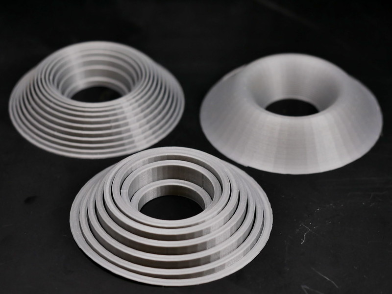
These printed models illustrate the Shell Integration method for estimating the volumes of solids of revolution . Professor Thiel uses these when teaching integration in Calculus II (MAT1575).
Cavalieri’s principle
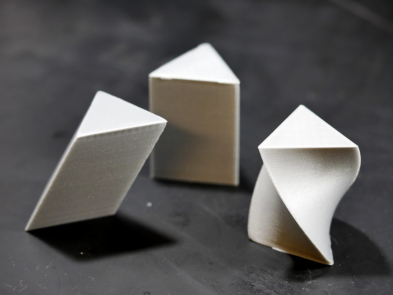
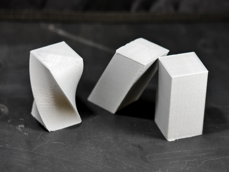
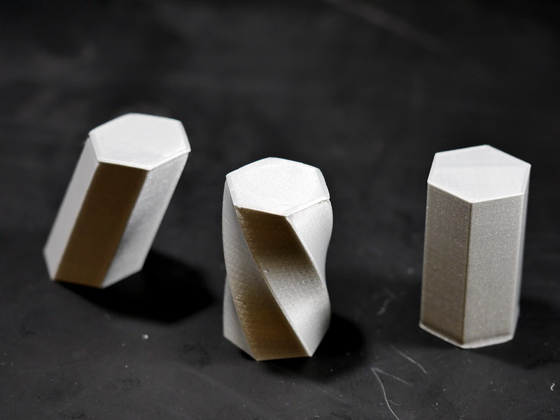
The following models illustrate Cavalieri’s Principle: any two solids of equal height with identical cross-sectional areas have the same volume. In each image below, all three of the printed shapes have the same volume.
Biological Models
The Ultimaker was borrowed by Professor Seto to explore the various types of models that could be printed for Biology pedagogy.
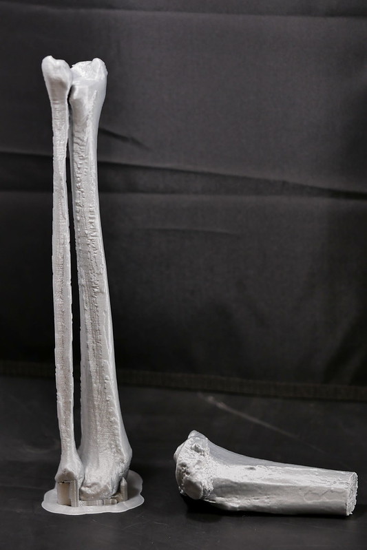
If Lucy fell… (L) Human fibula and tibia. (R) Fragment of tibia from Austrolopithecus afarensis
The first model attempted was a human fibula and tibia combination. This was generated as a comparison for the second model, a fragment of the tibia from the Austolopithecus afarensis called Lucy. The file for printing was provided from eLucy as a result of re-scanning for the Nature paper Perimortem fractures in Lucy suggest mortality from fall out of tall tree (Kappelman et al., 2016). These models were created to illustrate comparative anatomy and evolution and led to the failed printing of Hominini skulls.
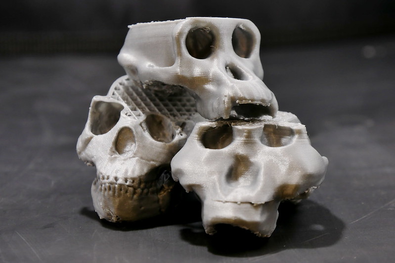
Partial Hominini skulls. Homo ergaster (bottom left), Paranthropus boisei (bottom right), Pan troglodytes (top)
Research
Smaller models were inspired by the personal research of Professor Chris Blair, an evolutionary biologist and herpetologist. Professor Seto identified an existing model of a horned lizard and assembled 2 additional models from existing CT-scans.
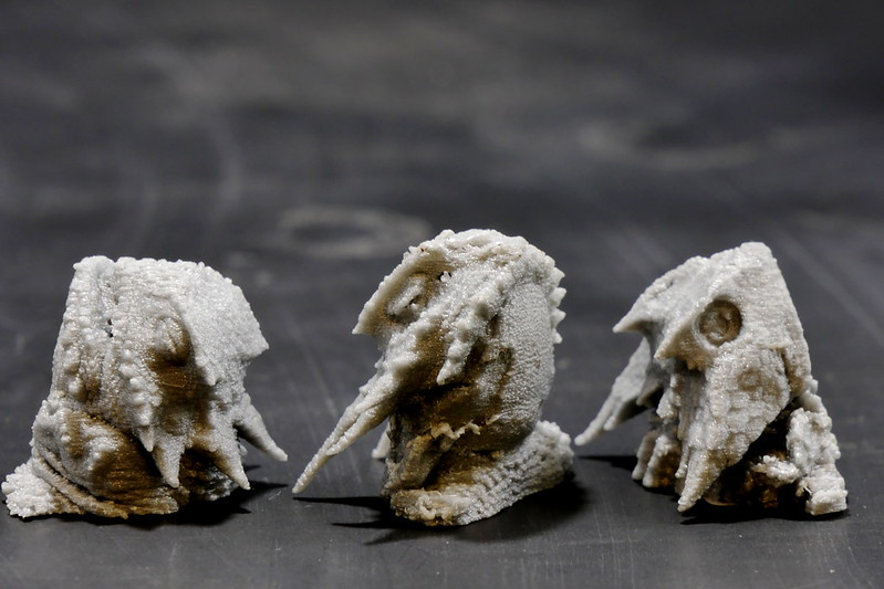
Desert horned lizard (Phrynosoma platyrhinos), Flat-tail horned lizard (Phrynosoma mcallii), Mexican horned lizard (Phrynosoma taurus)
Microbiology and Molecular Biology Pedagogy
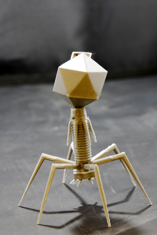
Assembled model from 3D printed parts of Bacteriophage T4
A multi-part model with assembly required was generated for a T4 bacteriophage for use in Molecular and Cell Biology (BIO3620) to illustrate the parts of the virus and the pivotal role in identifying DNA as the inherited genetic material.
Near Field Communication
Since models already exist in some classes, the idea to enhance the experience of learning through integration of technology was attempted using NFC to re-direct to existing videos or OpenLab sites about the model. In the future, Professor Seto hopes to work with faculty from other departments to augment the manipulatives with more complex interactions.
References
Kappelman J, Ketcham RA, Pearce S, Todd L, Akins W, Colbert MW, Feseha M, Maisano JA, Witzel A. Perimortem fractures in Lucy suggest mortality from fall out of tall tree. Nature. 2016 Sep 22;537(7621):503-507. doi: 10.1038/nature19332. Epub 2016 Aug 29.

