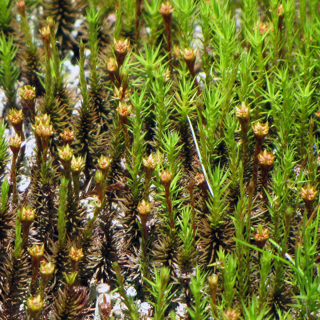
Polytrichum moss plants – male and female gametophytes – on Roan Mountain, Tennessee. Credit: BlueRidgeKitties via Flickr, CC BY-NC-SA 2.0 DEED
Station IIa
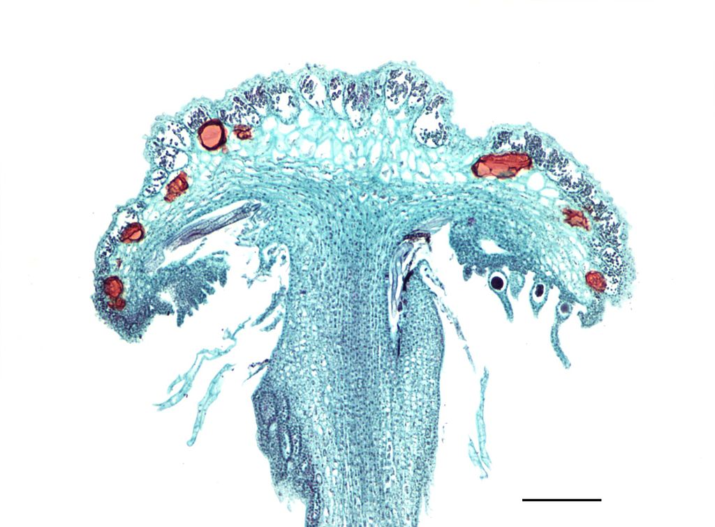
Longitudinal section of the archegonial receptacle of Marchantia. On the bottom surface can be seen the eggs within the venters, attached by the base of the archegonium with the necks pointing downwards. Scale = 0.2mm. Credit: Jon Houseman via Wikimedia, CC-BY-SA 3.0
Station IIb
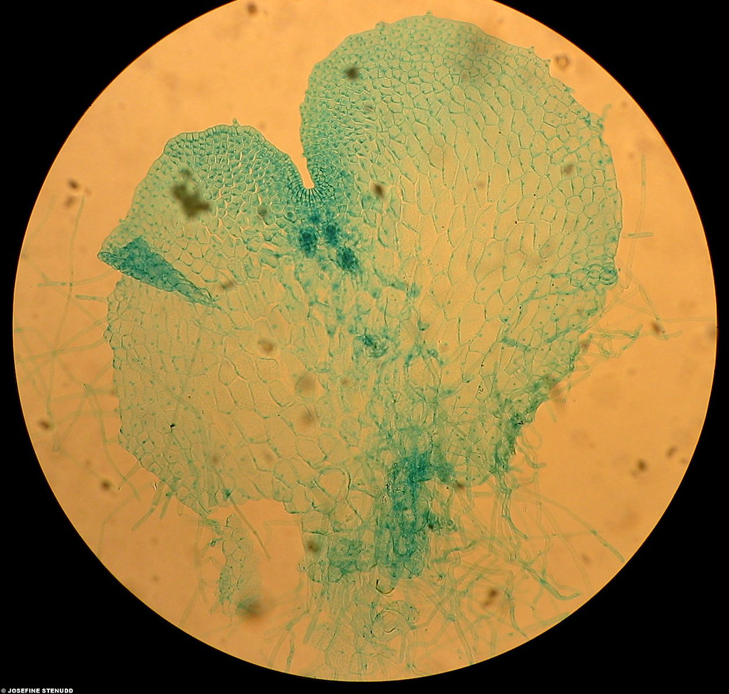
Bryophyta Mnium antheridium (red box) filled with flagellated sperm cells at the tip of a male gametophyte, seen under a microscope. Credit: Bruce Kirchoff via Flickr, CC BY 2.0 DEED
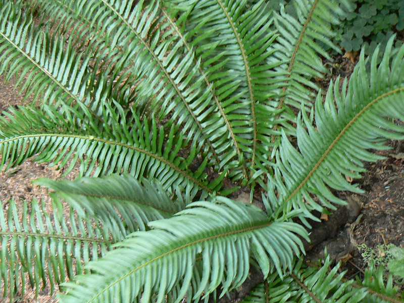
Station III
Sword Fern (Polystichum munitum) fronds. Credit: Willamette Biology via Flickr, CC BY-SA 2.0 DEED.
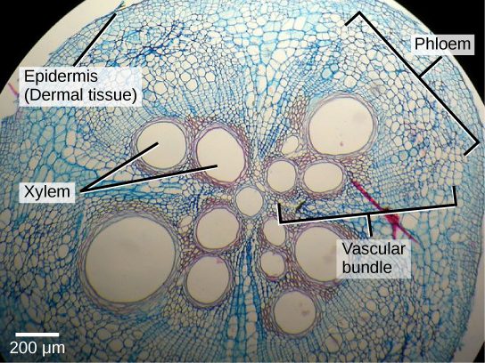
Station IVa
Light micrograph showing a cross section of a squash (Cucurbita maxima) stem. Each teardrop-shaped vascular bundle consists of large xylem vessels toward the inside and smaller phloem cells toward the outside. Xylem cells, which transport water and nutrients from the roots to the rest of the plant, are dead at functional maturity. Phloem cells, which transport sugars and other organic compounds from photosynthetic tissue to the rest of the plant, are living. The vascular bundles are encased in ground tissue and surrounded by dermal tissue. Credit: CNX OpenStax via Wikimedia, CC BY 4.0
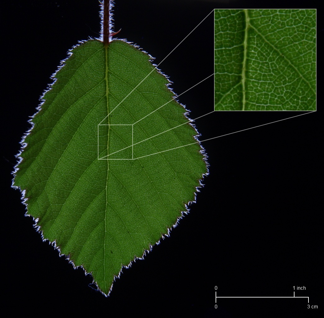
Station IVb
Detail of the vasculature of a bramble leaf. Credit: Zephyris via Wikipedia, CC BY-SA 3.0
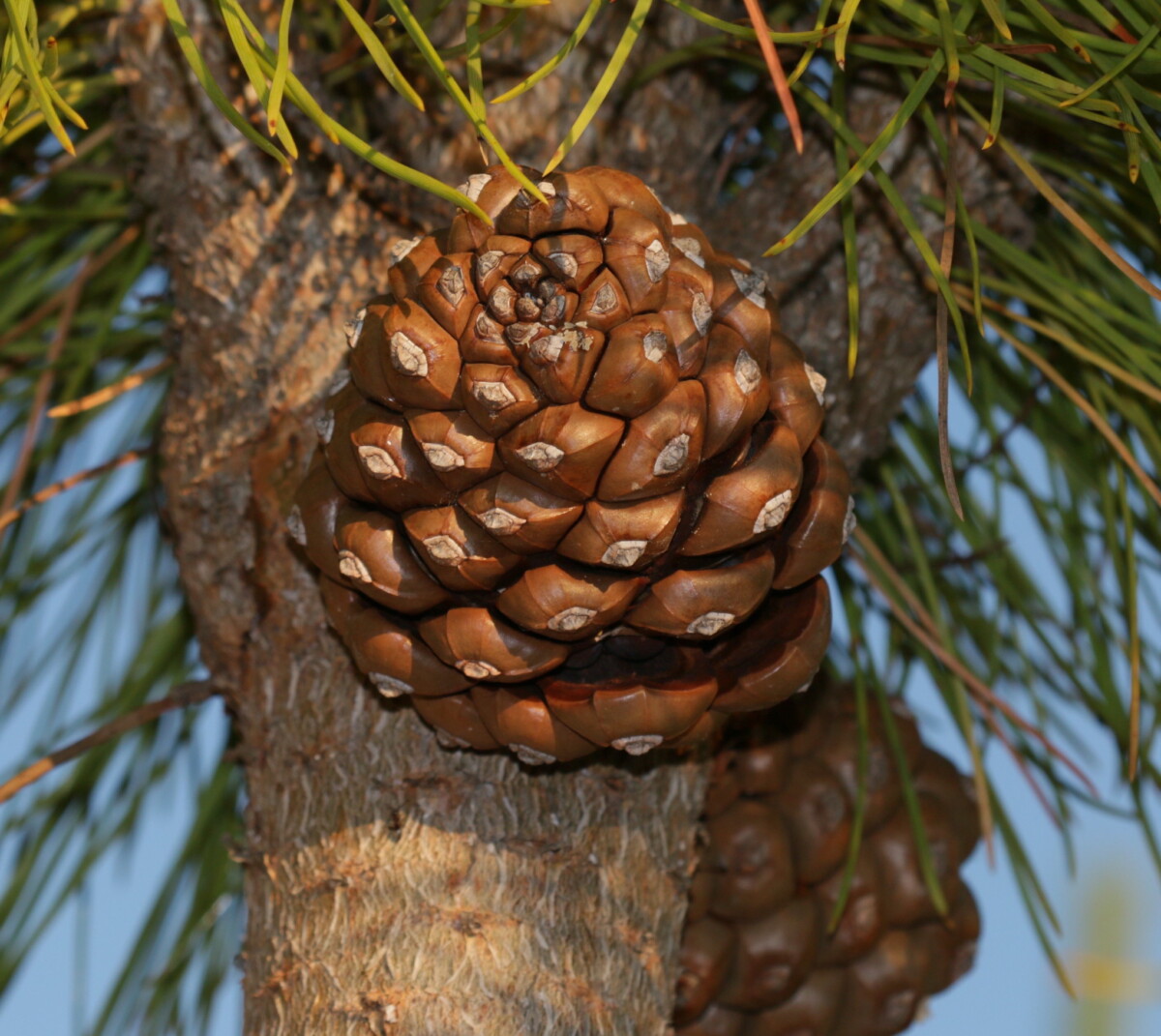
Station Va
Pinus pinea (Stone Pine) – cone. Credit: S. Rae via Flickr, CC BY 2.0 DEED
Station Vb
Cones of Stone Pine (Pinus pinea L.). Credit: Giancarlodessi via Wikimedia, CC BY BY-SA 3.0
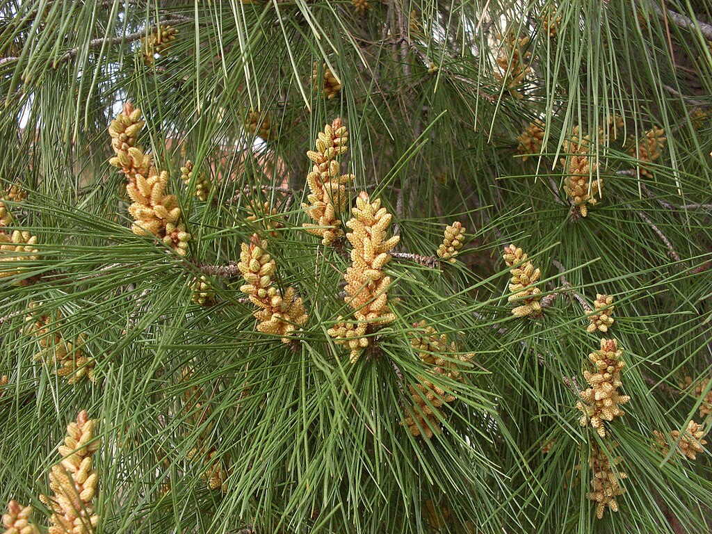
Station VI
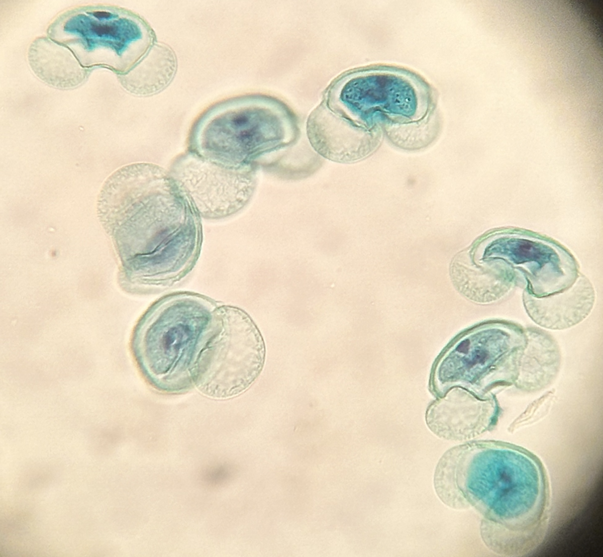
Stained pine pollen observed under a light microscope at 40X. Credit: Tatiana Voza, CC BY-SA 4.0
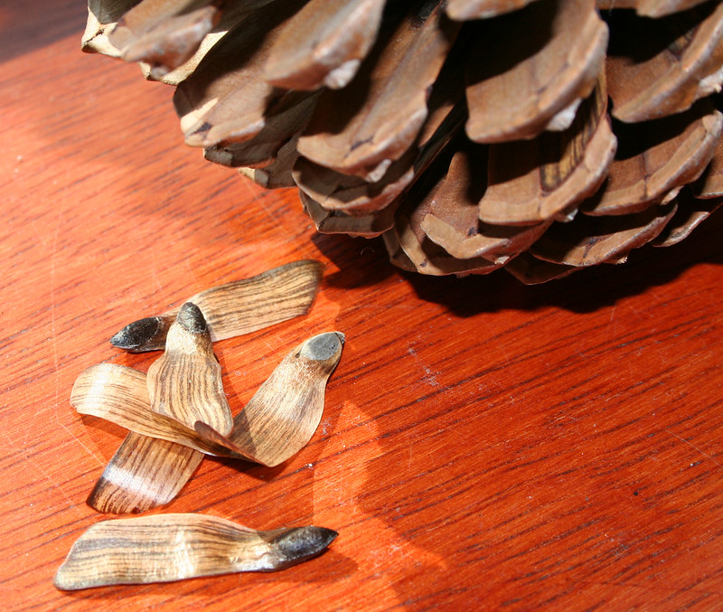
Station VII
A pine cone and some of the winged seeds that were in it. Credit: Frank Green via Flickr, CC BY-NC 2.0 DEED

