Laboratory III – The Fungi

1. Instructions

  • Open and download the worksheet.
  • Go over each station one by one
  • Answer the corresponding questions using the lab manual Laboratory III text

2. Worksheet

Open/Download/Print the Lab III worksheet before going over the stations  

3. Stations

Click on the image to enlarge it and see more details

Station I


translucent oval shaped cells of different sizes, some clustered together on a gray background
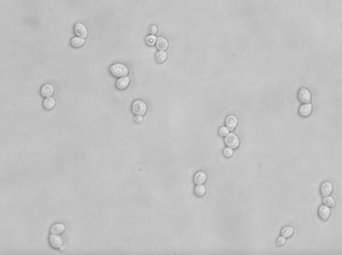
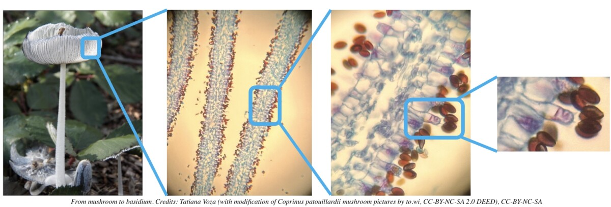
Still shot taken from fresh budding yeast (Saccharomyces cerevisiae) video, growing and, er, budding… 2 hour time-lapse. 2.5 mins per frame. 48 frames, played at 4 frames per second. Ligh microscope x400. Credit: Darren Wilkinson, CC BY-SA 2.0 DEED , via Flickr

Watch the video of budding yeast cells under the microscope x400

Station II

Pink branching tubular structures ending with several aligned small oval ones
Stained Penicillium conidia observed under the microscope with a x400 magnification. Credit: Tatiana Voza, CC-BY-NC-SA 4.0 DEED


A translucent and purple tube-like structure with a round end and many small purple circles arranged in rows surrounding the cercle.
Stained conidia of Aspergillus fumigatus observed with a microscope x400. Credit: Public Domain

This is what Penicillium and Aspergillus molds look like to the naked eye

Station III

Translucent elongated sac-like structures containing purple oval shapes
Asci from Peziza ampliata stained with Toluidine Blue observed with microscope at x1000 magnification. Credit: T, CC BY-NC-SA 2.0 DEED via Flickr

These are Peziza fungi observed with a naked eye

Station IV

Purple blue structures organized in layers with oval brown shapes clustered next to them
Light microscopy of stained Coprinus basidia. Credits: Tatiana Voza, CC-BY-NC-SA 4.0 DEED

See where the basidia are located on the fruiting body (mushroom)

Station V

Green filaments with a spherical end showing numerous small brown structures inside.
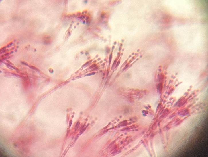
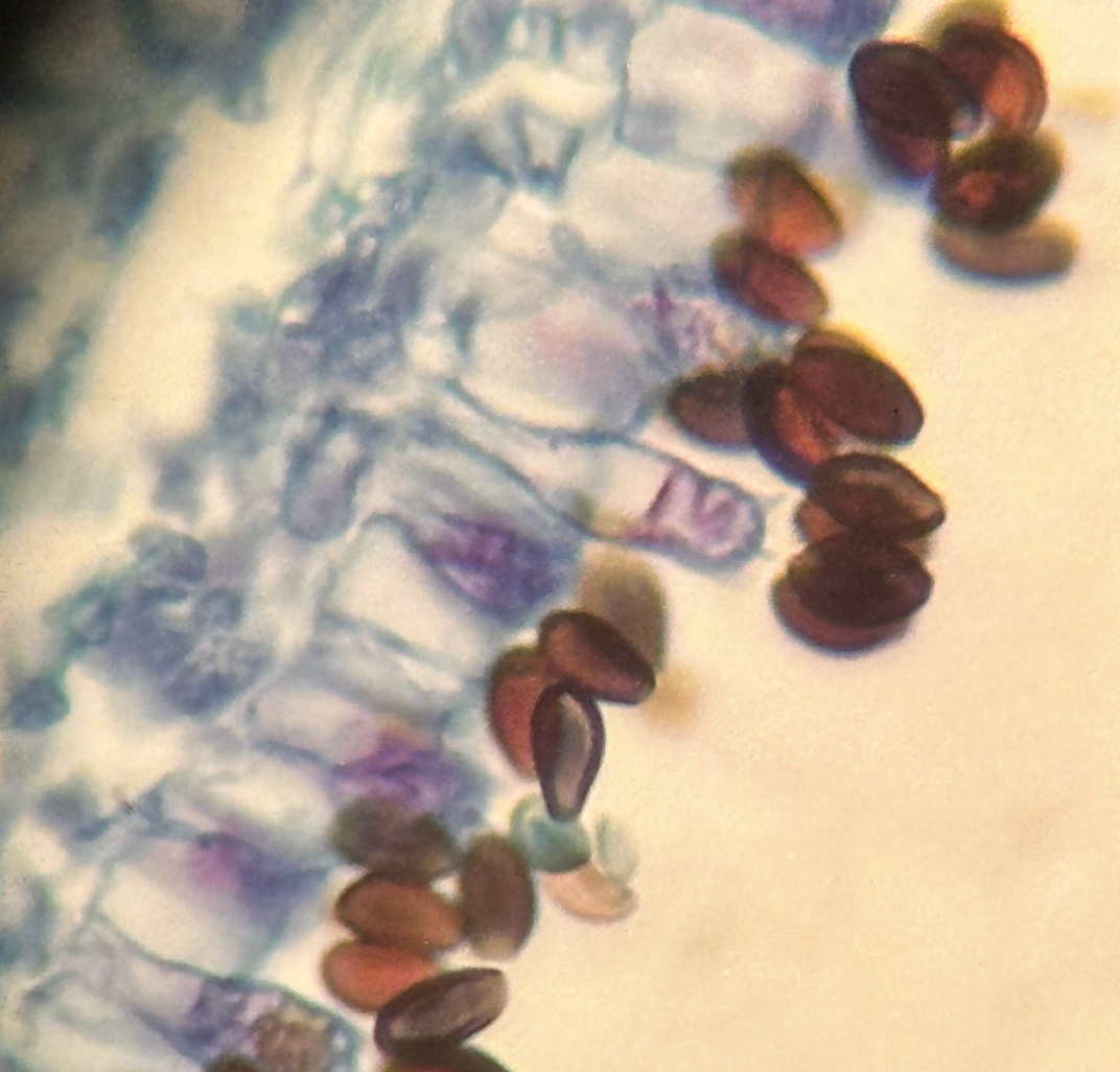
Light microscopy of stained Rhizopus showing a close up view of two sporangium of Rhizopus where the differentiation between the spores and columella can be seen attached to the hypha. Scale bar = 0.1mm. Credit: Jon Houseman, CC-BY-SA 3.0 via Wikimedia

See Rhizopus mold observed with a naked eye

Station VI

Green filaments with larger structures connecting around a black oval shape with jagged edges
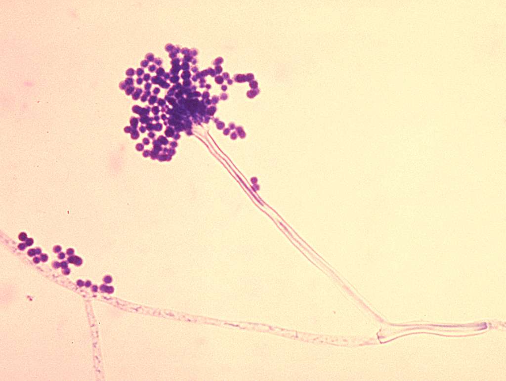
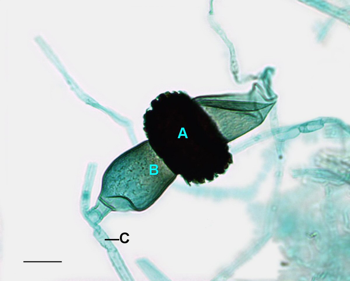
Light microscopy of stained Rhizopus showing a mature zygosporangium with what is believed to be the sprouting sporangiophore that would release the meiospores. A=Mature zygosporangia, B=Sprouting sporangiophore, C=Suspensor cell. Scale bar = 0.1mm. Credit: Jon Houseman, CC BY-SA 3.0 via Wikimedia


Station VI

Fresh oyster mushrooms. Credit: Marco Verch, CC-BY 2.0 DEED, via Flickr


Station VIII

White papery blob on dark reddish brown tree bark
Lichen on Manzanita tree bark in the Petrified Forest Park, Calistoga, California, USA. Credit: Tatiana Voza, CC-BY-NC-SA 4.0 DEED

Go Back to Top