1. Instructions
- Open and download the worksheet.
- Go over each station one by one
- Answer the corresponding questions using the lab manual Laboratory II text
2. Worksheet
Open/Download/Print the Lab II worksheet before going over the stations
3. Stations
Click on the image to enlarge it and see more details
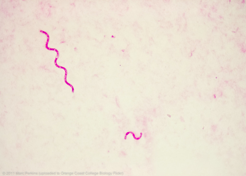
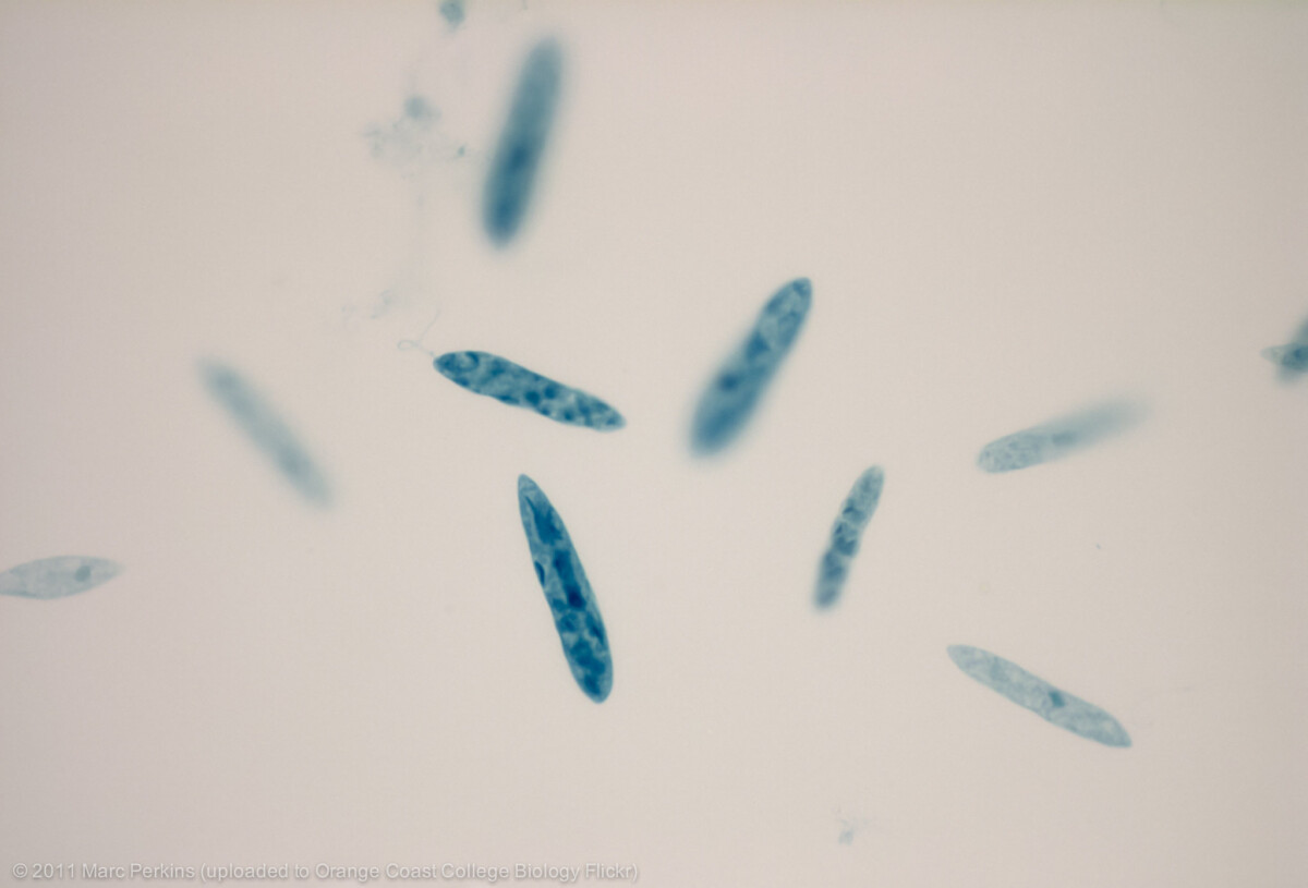
Station Ia
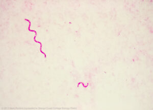 Microscopy slide of Gram-stained Spirillum volutans 1000x. Credit: Marc Perkins, CC-BY-NC
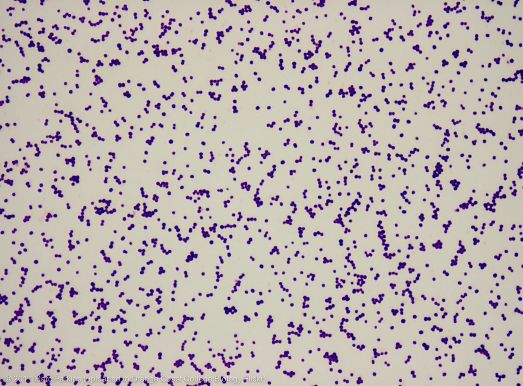
Station Ib
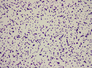 Microscopy slide of Gram-stained Staphylococcus epidermidis 1000x. Credit: Marc Perkins, CC-BY-NC
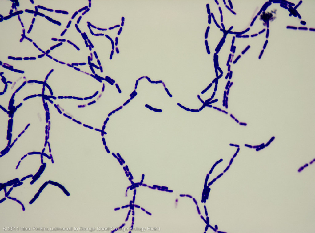
Station Ic
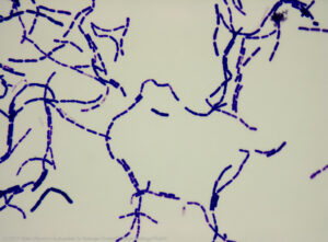 Microscopy slide of Gram-stained Bacillus megaterium 1000x. Credit: Marc Perkins, CC-BY-NC
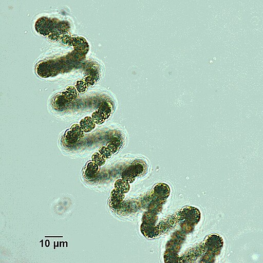
Station II
 Live cyanobacteria from genus Dolichospermum seen under a microscope. Credit: Masa Zupancic, CC-BY-SA 4.0
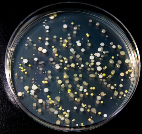
Station III
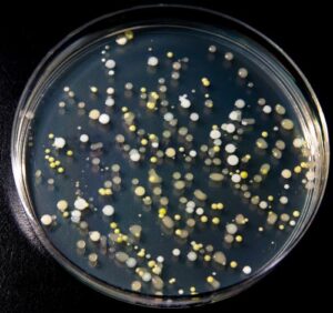 Microbial culture resulting from swabbing the surface of a cell phone. Credit: Carlos de Paz CC-BY-SA 2.0 DEED
Station IV
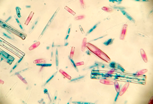 Various stained diatoms seen under a light microscope x400. Credit: Tatiana Voza, CC-BY-SA 4.0
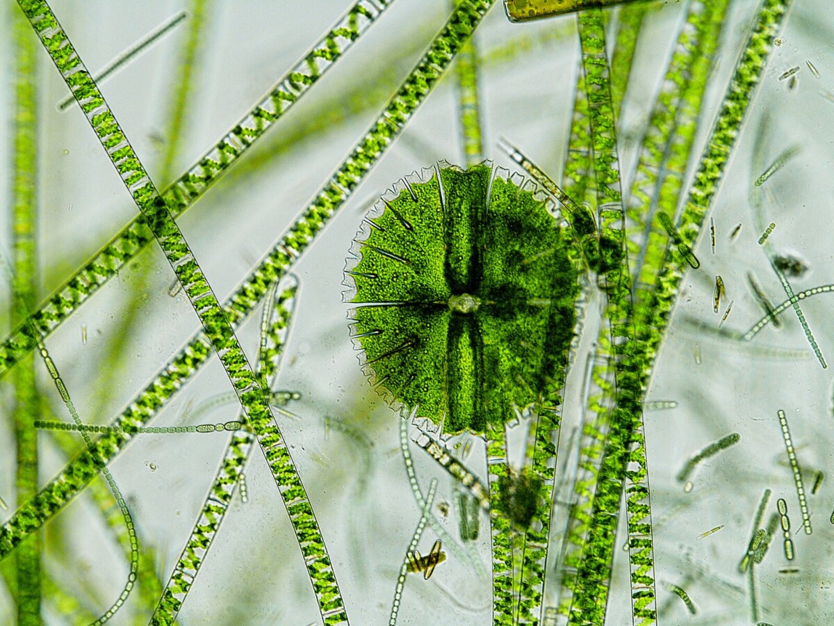
Station V
 Zygnematophyceae, Spirogyra, Micrasterias, algae, microphotography (x1000), environmental sample, fresh water, mountain stream, Jizerské Hory (Czech Republic, Liberec Region). Credit: Vosolsob, CC-BY-SA 4.0
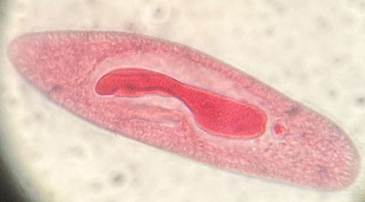
Station VI
 A stained paramecium observed under the microscope x400. Credit: Tatiana Voza, CC-BY SA 4.0.
Video of live paramecia under the microscope
Station VII
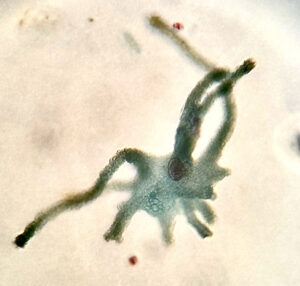 Stained amoeba observed under the microscope x400. Credit: Tatiana Voza, CC-BY SA 4.0
Station VIII
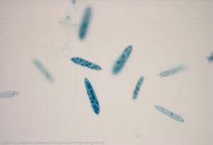 Stained euglena observed under the microscope x400. Credit: Marc Perkins, CC-BY NC 2.0 DEED
Video of live Euglena under the microscope
Back to Top