
Attached are photographs of interesting findings, that I have seen during my clinical experience.
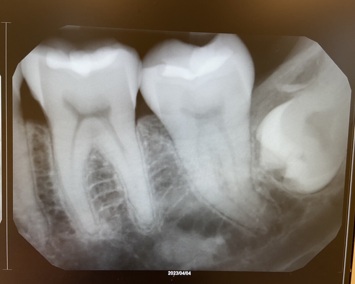
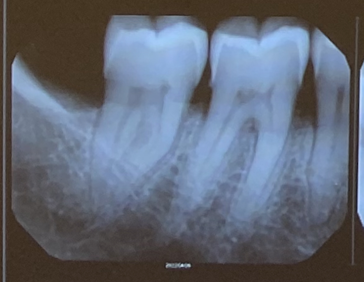
In the above PA image, a radiopacity can be seen inferior to teeth #18-#19, that could potentially be a salivary gland stone.
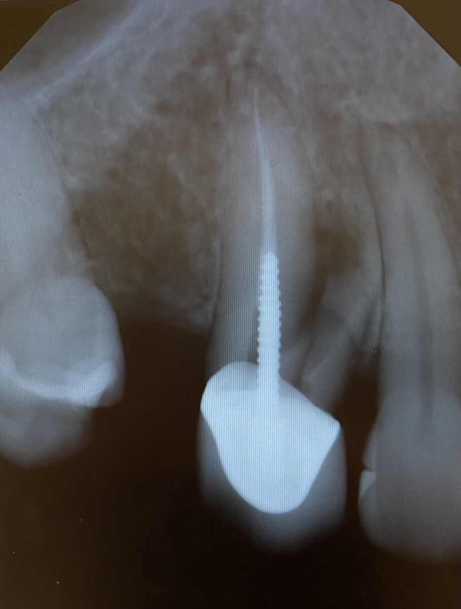
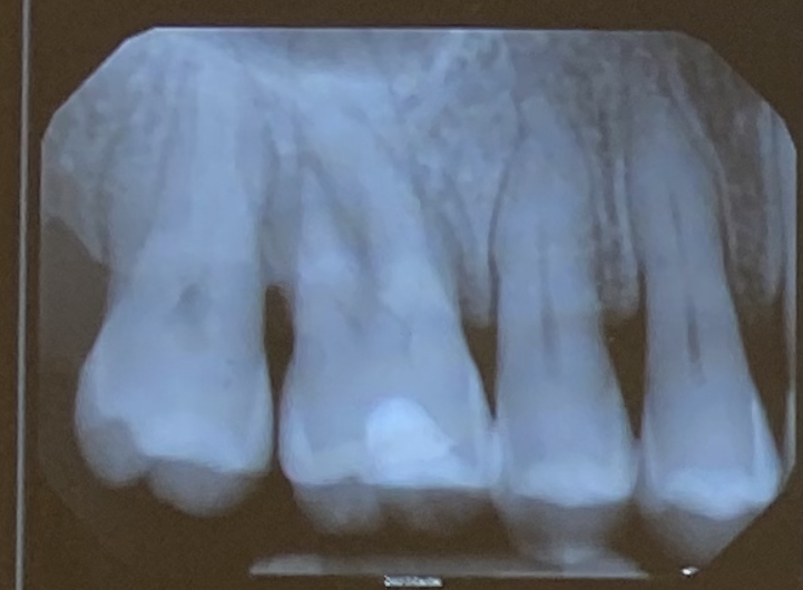
In the above PA image, a large root fracture can be seen on tooth #6.
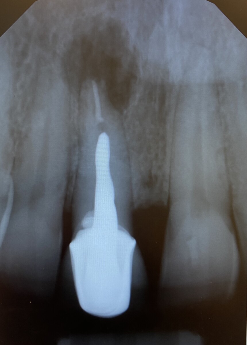
In the above PA, a large radiolucent area can be seen surrounding the apex of tooth #8
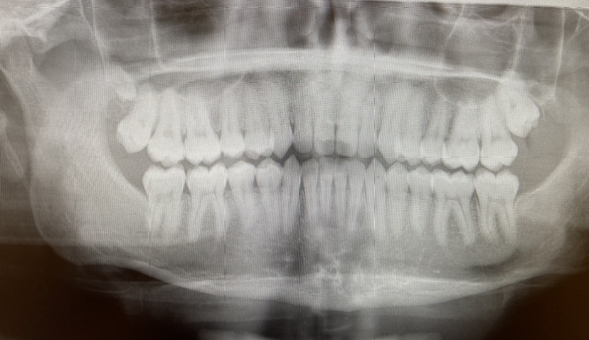
In the above PAN, a supernumerary tooth can be seen distal to tooth #1
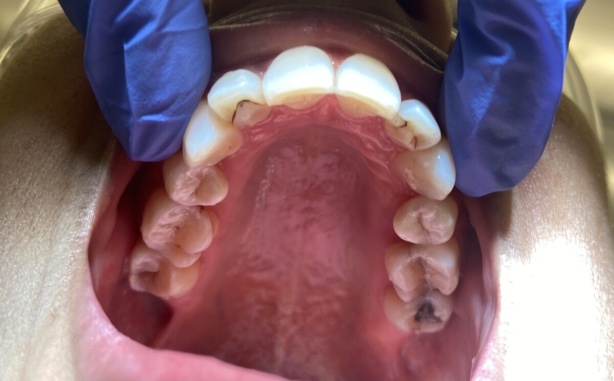
The photo above shows talon cusps present on teeth #7 and #10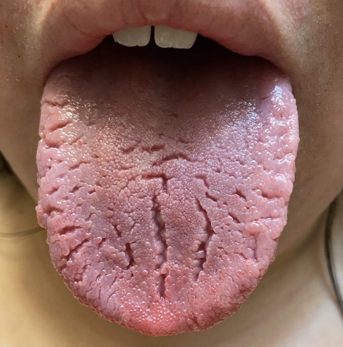
The photo above shows a fissured tongue
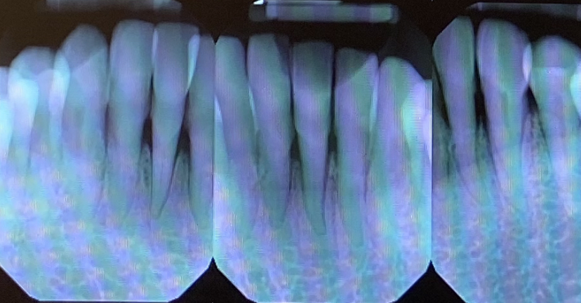
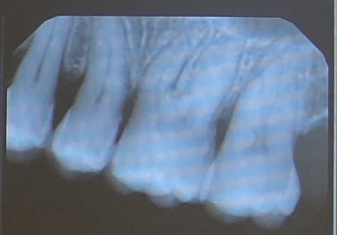
The 4 photos above depict an advanced periodontally involved case with furcation involvement (Stage III Grade C Periodontitis)

