CASE 1:
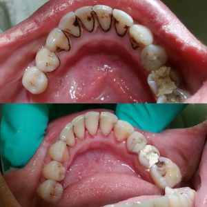
Patient S.K 36 year old Asian female. Patient has no medical condition, no allergies, not taking any medications, no hospitalizations. She has not had dental cleaning for 5 years H/II with severe BOP and generalized heavy supragingival and subgingival calculus deposits and heavy staining. Pocket depths 4-5mm, localized 6-7mm. No mobility. Gingival tissue is erythematous with moderated marginal and papillary inflammation. I completed all the assessments, educated the patient on proper tooth brushing (modified bass method) and proper flossing technique, full mouth scaling using ultrasonic and hand instruments, rubber cup polish with coarse paste and sodium fluoride varnish.
Case 2
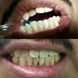
Patient S.K 42 year old Hispanic male. Patient has no medical condition, no allergies, not taking any medications, no hospitalizations. He has not had dental cleaning for 1 year H/II with moderated BOP and generalized heavy supragingival and subgingival calculus deposits. Pocket depths generalized 4-5mm, No mobility. Gingival tissue is moderated marginal and papillary inflammation. I completed all the assessments, educated the patient on proper tooth brushing (modified bass method) and proper flossing technique, full mouth scaling using ultrasonic and hand instruments, rubber cup polish with fine paste and sodium fluoride varnish.
Case 3
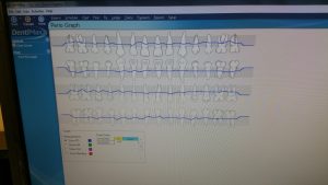
Patient: S.H Age 29, Asian Female, H/II. ASA I, Patient does not have any medical problem. NO systemic condition. NO medication. No hospitalization. No allergies. Pt had orthognathic surgery due to Class III occlusion. Class I occlusion and edge to edge bite anterior. Patient is classified as a heavy; localized supra-gingival calculus in anterior mandible and generalized maxillary and mandibular posterior subgingival calculus. Pale pink gingival tissue with generalized moderate marginal inflammation. Patient had moderate BUP; probing depths 1-6mm. Patient has Type II generalized periodontitis. Radiographs showed 15% horizontal bone loss in posterior mandible.
Based on initial assessment, patient has moderate inflammation, has pocket depths of 5 to 6mm and no allergies to any medication. The sites that are possible to be qualified for Arestin which are the following: tooth #18(DL,DB) with 5mm, #19(ML,DB) with 5mm, #20(ML) with 5mm, #21(DB, ML) both with 6mm, #22(ML) with 5mm, #23(DB) with 5mm, #26(ML) with 6mm, #28(DL) with 6mm, #29(DL) with 6mm, #30(DL) with 6mm, and #31(ML) with 5mm. After week, evaluated LRQ and LLQ due to place to Arestin; the gingiva tissue is pink, less inflamed, film and improved tissue adaptation. Re-probing #18-23 and #26-31. The probe depth are #29(ML,DL) with 5mm, #30(ML) with 5mm, (DL) with 6mm, and #31(ML) with 6mm. I was planning to place Arestin both side mandibular; however, when patient came back after scaling, her probe depth reduced and gingiva was firm and well adapted to the teeth. I placed Arestin five sites which were #29(ML,DL) with 5mm, #30(ML) with 5mm, (DL) with 6mm, and #31(ML) with 6mm. After 6 week, pt’s pockek depth reduced.
| TOOTH # | SITE | BEFORE | ATRET | RATE |
| 29 | ML | 5 | 4 | 20% |
| 29 | DL | 5 | 4 | 20% |
| 30 | ML | 5 | 3 | 40% |
| 30 | DL | 6 | 3 | 50% |
| 31 | ML | 6 | 4 | 33.3% |


