
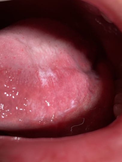
This patient was a 24-year-old Asian male with an ASA II classification. Based on the perio exam and radiographs they were identified as Perio Stage II Grade C. During the intra-oral exam a white patch (leukoplakia) was discovered on the ventral side left side of the tongue. The patient was not aware of the lesion. Initially, gauze was used to possibly remove the superficial layer of the mucosa, however, the lesion remained. This indicated to me that it was indeed a lesion and not sloughing of the oral mucosa. Since the patient was a tobacco smoker it was imperative to counsel the patient on smoking cession. The patient was very concerned, and it was explained to the patient that it could be caused by a myriad of things. It could be something benign such as hyperkeratinization or malignant as cancer. The lesion was measured (3mm h/5mm w) and the patient was given a referral for oral pathology. The patient was told they must get a biopsy of the lesion to determine its classification.
During the second and final visit, the patient reported that they did not go to the doctor, but they made an appointment to see one the following week. Also, to my surprise, the patient revealed that they decided to stop smoking. The lesion was measured again with no changes in size. It was then reiterated to the patient the importance of smoking cessation and getting a biopsy of leukoplakia on their tongue.

