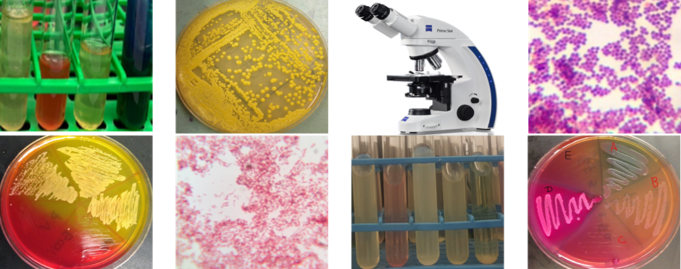
Results from Schaeffer-Fulton method of endospore staining using B. subtilis
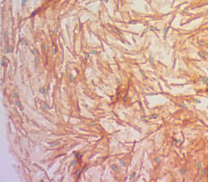
Figure 1. Endospore stain using B. subtilis
Figure 2. Endospore stain using B. subtilis
Questions
- Describe the results shown in figure 1 and 2.
- Which of the above figures shows stain from a) less than one week, b) more than one week? Explain your choices.
- What type of endospores (in terms of location of the spores) are produced by B. subtilis?
- Compare and contrast endospore stain, acid-fast stain and Gram stain. Be sure to include all similarities and differences.
- Why are some bacteria producing endospores likely more virulent than those not producing endospores?
- Name three other virulent factors found in or associated with other bacteria.
Negative Stain

Figure 1. E. faecalis
Figure 2. B. subtilis
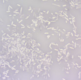
Figure 3. P. aeruginosa
Questions
1.Why are these called negative stains?
2.Describe the shape of each of these bacteria?
3.Is it possible to tell if these bacteria in figures 1 – 3 produce capsules? Why or why not?
4.What further step could have been taken to make capsules more visible?
5. How do capsules add to bacteria’s virulence? Why?

