Case #1:
- Demographics:
- 46 years old
- White
- Male
- Assessments:
- Patient has pre-existing conditions of high blood pressure and dizziness. The patient is taking the following medications on the daily basis: Meclizine – 25mg, Ramipril – 5mg, Omega 3 – 1000 mg, no recent hospitalizations, and no allergies. Vital signs within normal limits. Smoker. ASA II.
- Patients last dental visit was in June 2016, had an exam, dental radiographs, and cleaning done. Patients last medical exam was January 2021.
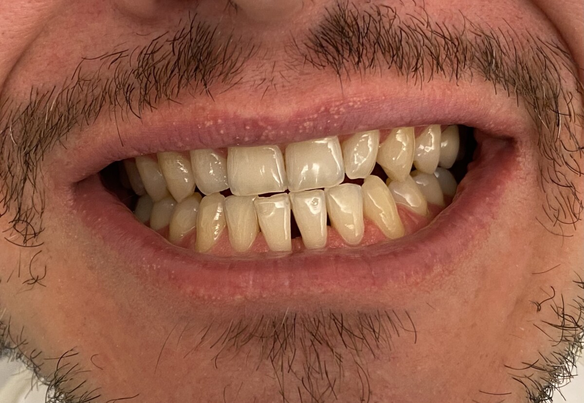
- Extra Oral findings: right side, small, round, movable submandibular lymph nodes -asymptomatic. Intra Oral findings: Moderately coated tongue. Right side 2mm macule (beauty mark) localized on the right side by postauricular.
- Dental Assessment: Class of occlusion right side class I, left side class II. Overjet 12mm and overbite 80%. Attrition: #21-27 and #6-12.Dental restorations have been noted on teeth #2;#14;#15;#17;#30 and #31. Impacted #32. Suspicious carious lesions noted on #1;#12 and #19.
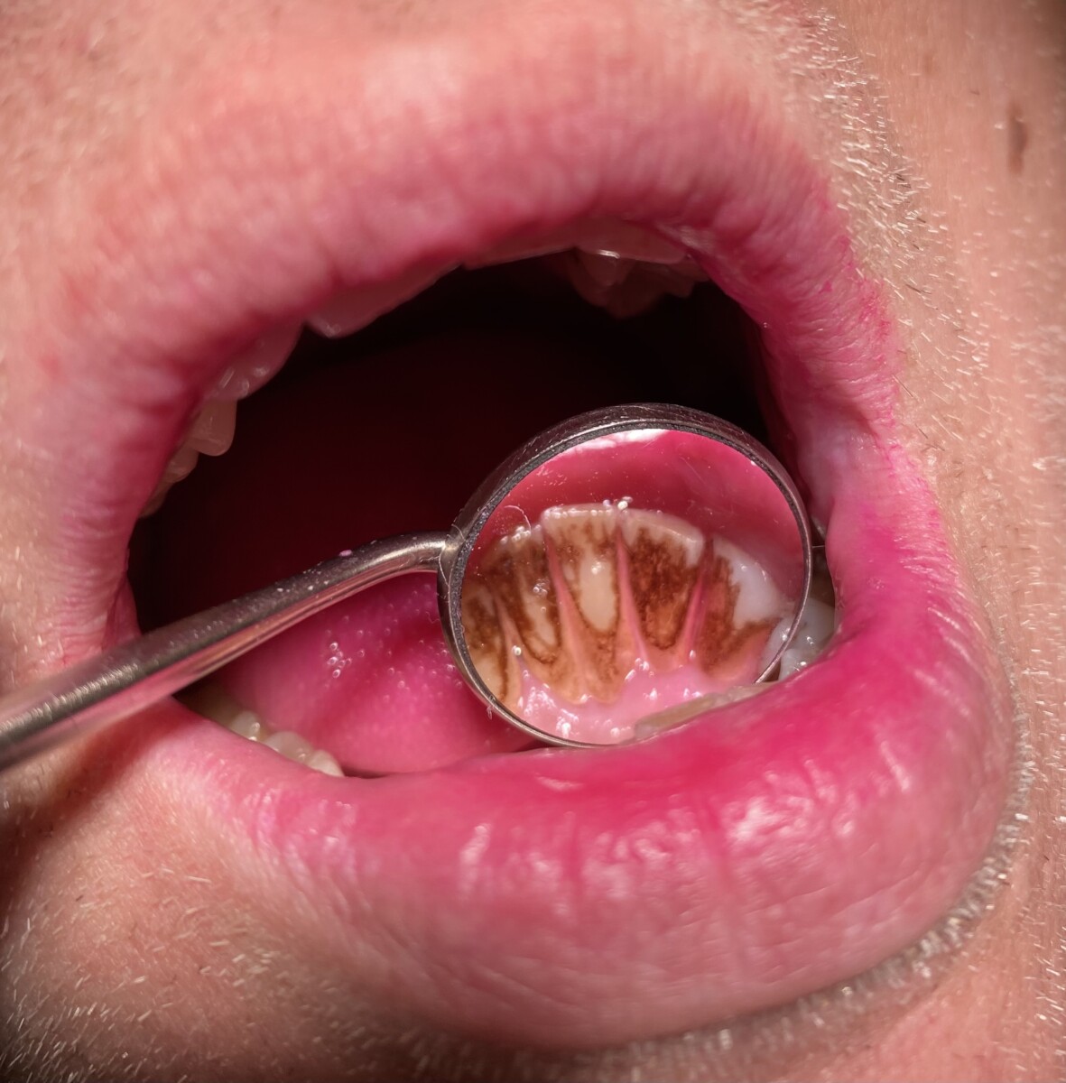
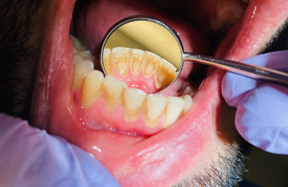
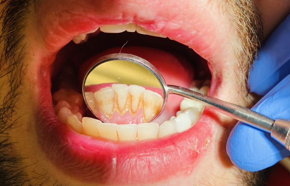
- Gingival tissue has generalized red inflamed gingiva, bulbous and spongy gingiva, blunted interdental papilla.
- Due to generalized supragingival and subgingival calculus deposits, probing pocket depths of 4-5 mm with moderate bleeding upon probing case value was determined to be heavy. Periodontal Stage II Grade B, due to localized 50% bone loss, generalized 35% bone loss, and localized CAL area of 4-5mm present.
- Planning and Implementation.
- Exposed FMX radiographs.
- Radiographic findings: Generalized bone loss, generalized interproximal visible. No interproximal caries visible, no PAP visible.
- Referral to DDS given to the patient for suspicious carries on teeth #1, #12, and #19.
- Full mouth scaling and root planing using hand instruments.
- Full mouth scaling and root planning using hand instruments completed in two visits.
- Engine polishing with medium grit prophy paste.
- Applied 5% NaF varnish, post-op instructions given to the patient.
- Oral Hygiene Instructions:
- Patient was taught modified Bass brushing technique to reduce the accumulation of plaque build-up. Patient was able to demonstrate proper technique back.
- Taught patient proper flossing technique. Advised to use water pick.
- Exposed FMX radiographs.
- Evaluation:
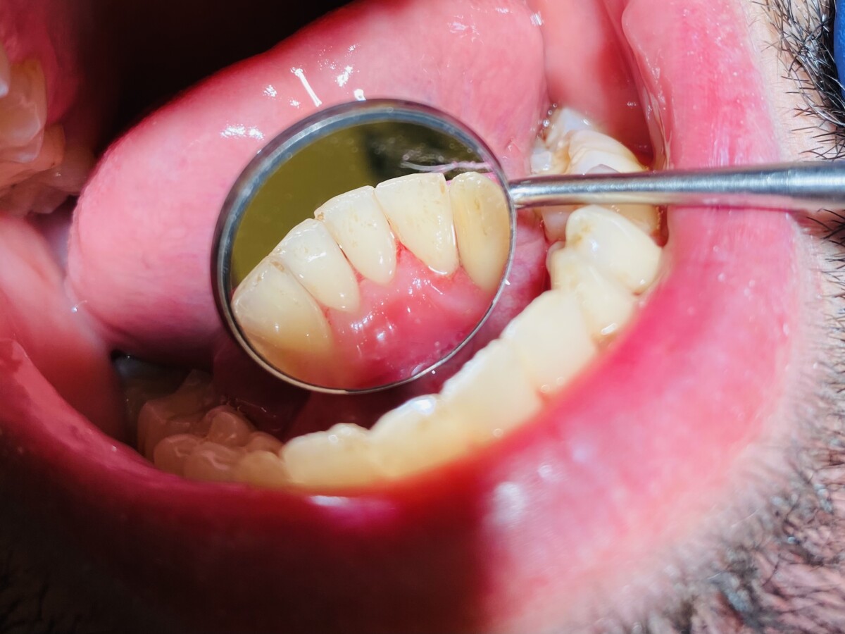
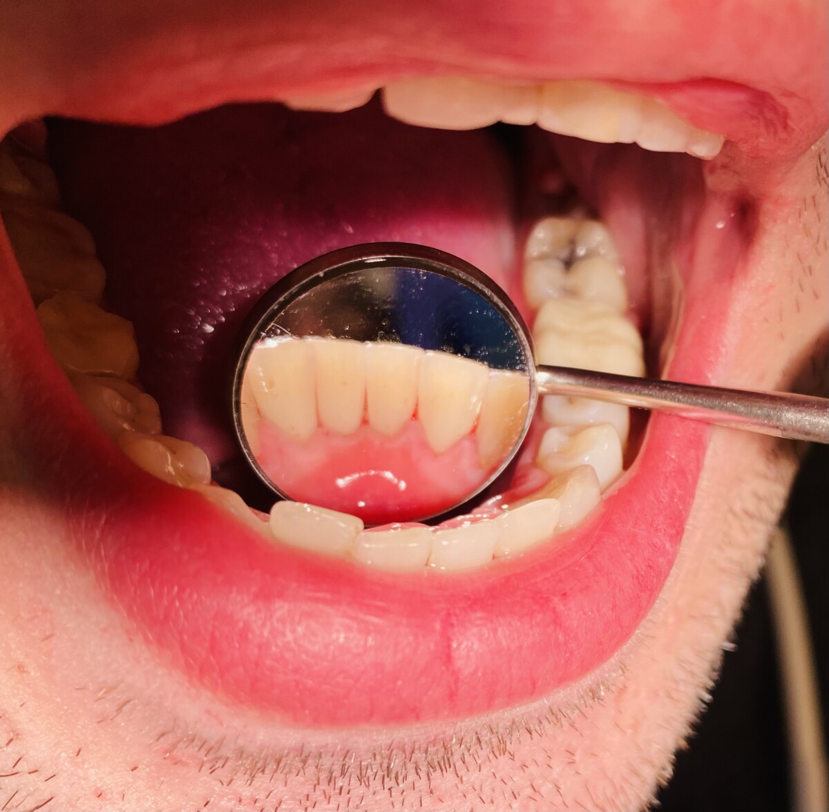
- Treatment was performed in two visits. On the second visit the gingival tissue of the previously treated area had less inflammation present and had responded well to treatment. Less bleeding was noted. Patient was advised 3 month recare appointment.
Before : After :
Case #2:
- Demographics:
- 69 years old
- White
- Female
- Assessments:
- Patient had pre-exsiting conditions of high blood pressure, high cholesterol and diabetes. The patient was taking the following medications: Labetalol – 200mg x 3 day (HBP), Edarbyclor – 25mg x 1day (HBP), Metformin – 1000 mg x 2 day (diabetes), Cresror 20mg x 1 day (high cholesterol), no recent hospitalizations, and no allergies. Patient has taken all her medications before the visit and have been taking them for the past ten years. Vital signs within normal limits. Smoker. ASA III.
- Patients last dental visit was in January 2016, had an exam, dental radiographs, and cleaning done. Patients last medical exam was February 2021.
- Extra Oral / Intra Oral: WNL
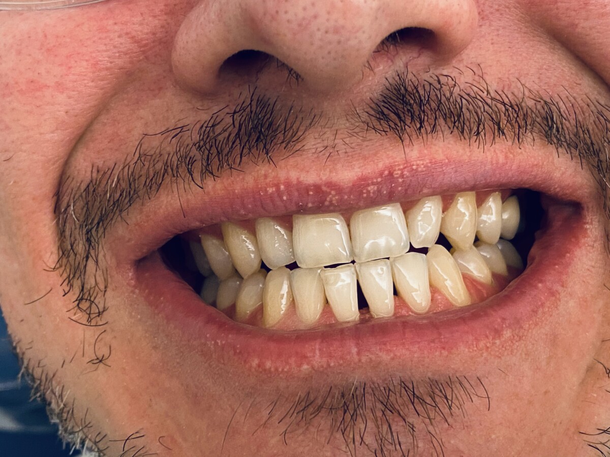
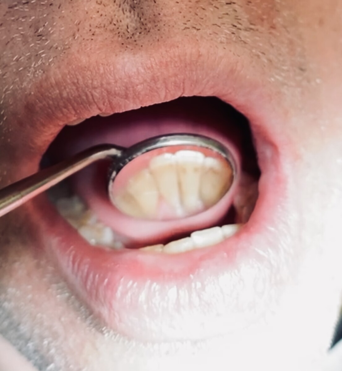
- Dental Assessment: Class of occlusion bilateral class I. Overjet 3 mm and overbite 20%. Generalized attrition on maxillary anterior teeth present. Abfraction present on #22;#8;#9;#10 and #11.Generalized dental restorations present. Cantilever bridge X – 4 -5. Moderate caries risk level due to exposed root surfaces. Caries lesion present on root #18.
- Gingival tissue has generalized pink color and localized inflammation to posterior teeth and blunted interdental papilla.
- Generalized probing pocket depths of 4-5 mm with mild bleeding upon probing and localized 7-8 mm probing depth on left maxillary posterior teeth. Due to generalized supragingival and subgingival calculus deposits case value was determined to be heavy. Periodontal stage: localized stage III Grade C due to localized 65% bone loss, generalized 35% bone loss. Clinical attachment loss was marked as localized 5-6 mm and generalized 2-3 mm. Class I mobility present on maxillary posterior teeth.
- Planning and Implementation.
- Exposed FMX radiographs.
- Radiographic findings: Generalized visible calculus. Generalized bone loss present. Retained root tip in arear of #2. No PAP visible. Patient was given referral to DDS for evaluation of existing restorations, and periodontitis for bone loss evaluation.
- Full mouth scaling and root planning using hand instruments completed in two visits.
- Engine polishing with medium grit prophy paste.
- Applied 5% NaF varnish, post-op instructions given to the patient.
- Oral Hygiene Instructions:
- Patient was taught modified Bass brushing technique to reduce the accumulation of plaque build-up. Patient was able to demonstrate proper technique back.
- Taught patient proper flossing technique. Advised to use a soft pick or water pick and power toothbrush.
- Exposed FMX radiographs.
- Evaluation:
- Treatment was performed in two visits. On the second visit, the gingival tissue of the previously treated area had less inflammation present and had responded well to treatment. Less bleeding was noted. Patient was advised 3 month recare appointment.
Some other cases of before and after:
#1:
Before: After:
#2:
Before: After:
Whitening :