Tobacco, Alcohol, and Stage 1 Hypertension
Initial Visit:
CC: “Exam and cleaning”
Demographic: 28 y/o Caucasian male, Blood Pressure: 139/89, Pulse: 64.
Medical History: Last physician visit was in 2022, patient reports bloodwork and annual physical as normal. Allergy to pet dander and pollen, takes Allegra as needed. Patient also reports taking Glusomaine 1x/day for joint support. Patient reports smoking 2-3 cigarettes a day and consuming 4-5 alcoholic beverages 2x/month. Patient is interested in quitting smoking.
ASA 2 due to cigarette smoking, alcohol consumption, allergies, and stage 1 hypertension.
Dental History: Last dental cleaning and exam: 2022. Patient brushes 2x/day using a manual toothbrush and flosses 2-3x/week. Patient reports occasional discomfort in LR and LL in 3rd molar area and has never had his wisdom teeth extracted.
Clinical Examination:
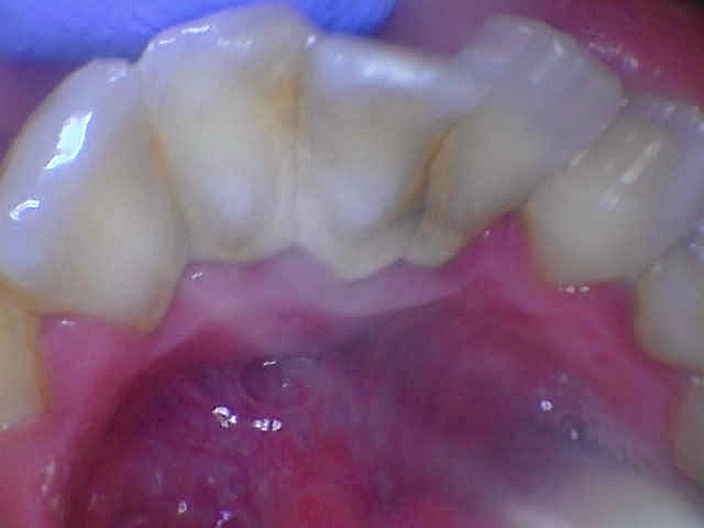
EO/IO: B/L discoloration on buccal mucosa (due to smoking), red linear burnt gingiva on lingual of #3 (pt reports burning gingiva eating hot food), tissue tag on upper labial frenum, tissue on left tonsilar area, and coated tongue.
Dental Charting: 2mm overjet, 95% overbite. Incisal fracture on #27. Anterior crowding present. #17 and #32 not clinically present.
Gingival Statement: Gingiva is highly inflamed, erythematous, rolled, and bulbous, with blunted papillae. Gingiva is swollen with a smooth and shiny texture. Gingival margin is 1-2mm coronal to the CEJ. Moderate bleeding upon gentle exploring.
This clinical presentation is consistent with smoking coupled with heavy supragingival and subgingival deposits due to lack of dental cleaning for over 2 years.
Periodontal Charting: Generalized 2-4mm with localized 5-6mm in posterior molar area. Moderate BOP.
-
- Calculus: Generalized supragingival and subgingival deposits; Case value: Heavy; Stain: Moderate
- Radiographic Statement: Panoramic imaging and 4 HBWs were consistent with less than 10% horizontal RBL, impacted #17 and #32, and generalized interproximal calculus present. No radiographic caries present. Patient informed of all findings.
- Periodontal Status: Generalized moderate gingivitis
- Caries risk: Moderate
- PI Score: 1.5- mainly interproximal and marginal
Reviewed medical and dental history, all assessments completed: EO/IO, dental charting, periodontal charting, calculus detection. Exposed PAN and 4HBWs. Provided patient with adult referral for evaluation of impacted 3rd molars and evaluation of tissue near tonsil area. OHI: Demonstrated c-shaped string floss to minimize interproximal plaque and explained the importance of flossing daily. Recommended an electric toothbrush (Philips Sonicare) to patient. Patient demonstrates understanding. Intraoral photos taken. Administered 0.5 cc of Oraqix in UR quadrant with patient consent; patient responded well. Scaled Q1 using ultrasonic and hand scaling.
Revisit #1:
Reviewed medical and dental history: F/u on adult referral– Pt has yet to schedule an appointment with medical provider. Pt reports using string floss as instructed every night. OHI: Reviewed string floss and demonstrated modified Bass brushing method to effectively minimize marginal plaque. Recommended a tongue scraper. Patient demonstrates understanding. Administered 1cc Oraqix in UL, LL, LR quadrants. Patient responded well. Completed scaling Q2, Q3, and Q4 using ultrasonic and hand scaling. Probed the linguals of the lower anterior teeth. Intraoral photos taken. Engine polished with medium paste, flossed all contacts, and applied 5% fluoride varnish. Post-op instructions given.
Recare: 3 months
In order to maintain care for the patient, a comprehensive approach considering medical, social, and dental aspects were considered. I first addressed and stressed the importance of yearly physical examinations. Since the patient last had his annual physical back in 2022, he was unaware of his stage 1 hypertension. The second being smoking cessation. Because the patient expressed interest in quitting, he had questions about the effects of smoking on his overall health. We discussed the implications of cigarette smoking in the oral cavity and its associations with oral cancer (and its increased risk when coupled with alcohol), decay, and periodontal disease, as well as the link between smoking and increased blood pressure. The patient presented with a tissue growth near the tonsilar area and impaction of 3rd molars, these were referred out to specialists for evaluation. Given the patient’s history of alcohol and tobacco usage coupled with heavy calculus build up, it is imperative to schedule more frequent visits every 3 months, providing intensive oral hygiene instructions such as flossing coupled with the modified Bass brushing method, and offering resources such as smoking cessation and purchasing an electric toothbrush. Implementing these methods in conjunction with nonsurgical periodontal therapy (SRP) will address persistent calculus build up every 3 months until the patient can demonstrate improved compliance in home care with decreased build up.
By addressing medical, social, and dental aspects along with comprehensive dental prophylaxis and regular monitoring, we can effectively manage the patient’s oral health and provide adequate care to prevent any future complications.
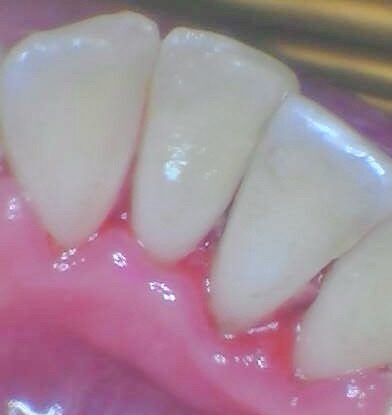
Before and After: