Case Study #1
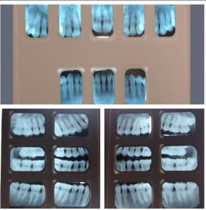
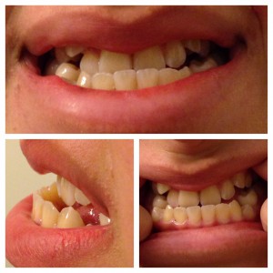
24 year old male, non-smoker, generally in good health, came into the clinic stating he had never been to a dentist for a check-up or had a cleaning. Patient presented with heavy calculus and staining. He had Type III Perio with generalized 3-7 mm pockets with severe bleeding upon probing. His gingiva was red, flaccid, with severe generalized inflammation. Upon taking a full mouth series of x-rays, there was radiographic calculus and bone loss noted. This patient was one of the few difficult cases I had throughout this program, but I was ready for the challenge. During review of homecare, the patient stated that he never flossed or rinsed and only brushed once a day, so meticulous oral hygiene instructions were given. The treatment plan was divided into 3 visits. The first visit was for scaling and root planning of the UR and LR quadrants, the 2nd visit was for scaling and root planning of the UL and LL quadrants, and the 3rd visit was to re-evaluate the quadrants and placement of Arestin. The patient was very concerned about his oral health and was glad he came to the clinic. He always wondered why his gums would bleed when eating an apple, but after explaining to him the importance of plaque removal , he understood why his gingiva was always inflamed and bled. When the patient returned 6 weeks after the placement of Arestin, both him and I were very impressed with the outcome. His gingiva was less inflamed, there was less bleeding, he was brushing twice daily, and flossing every night. The probe readings went down 2-3 mm in the area where Arestin was placed. Overall, the case was a success. I was proud of both the patient and myself. A referral was given to see the periodontist, and the patient was placed on a 3 month recall.
| Tooth number and surface | Initial probe readings | Post-arestin readings |
| 5 ML | 7 | 5 |
| 5 MB | 7 | 5 |
| 6 ML | 6 | 4 |
| 6 DL | 7 | 5 |
| 6 DB | 7 | 4 |
| 7 DL | 6 | 3 |
| 29 DL | 6 | 4 |
| 30 ML | 5 | 5 |
Case Study #2
I remember like it was yesterday. Histology/Embryology class during my first semester of the dental hygiene program, where I was chosen to give a presentation on Ectodermal Dysplasia. Ectodermal Dysplasia is a rare group of inherited disorders characterized by dysplasia of tissues of ectodermal origin, such as teeth, hair, nails, and skin. Dental manifestations include hypodontia and oral rehabilitation.
Fast forward to my last semester in the dental hygiene program, where I met my first patient with Ectodermal Dysplasia. 15 year old hispanic female, presented with a class III occlusion, and 9 missing teeth which included her premolars and molars. Patient was doing a good job with home care, so a light hand scaling was done followed by polishing. While speaking with the patients mom, I realized how much my patient went through while growing up. She was a strong girl and I was honored to see her as my patient. Her mother explained that the following year her daughter will be getting a mandibular osteotomy followed by rhinoplasty to correct her jaw alignment. Her mother told me that she herself had the same procedures done when she was a young girl and now she will watch her daughter do the same thing. The mother showed me a picture of herself before her surgery’s; while she stood in front of me and seeing the after, I was just blown away. I was amazed at the work that health professions can do and the impact it has on peoples lives. At that moment I realized why I chose to be in this profession. To see people smile, to give them support and keep them going!