Case 1
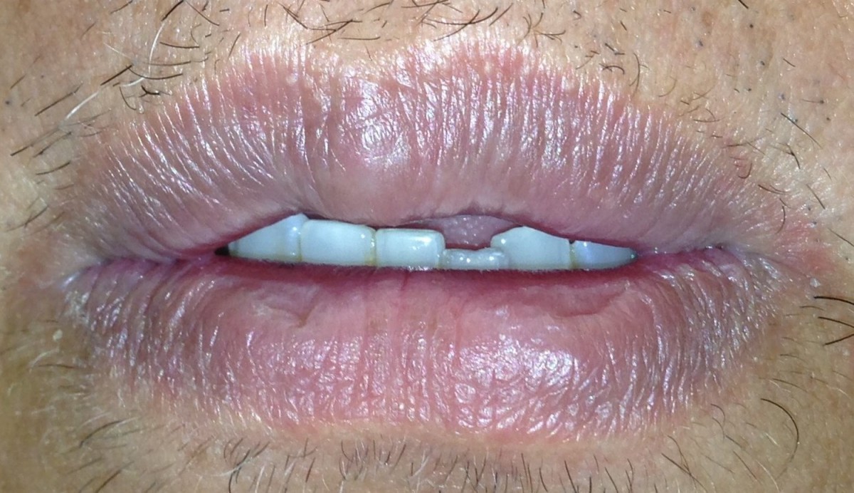
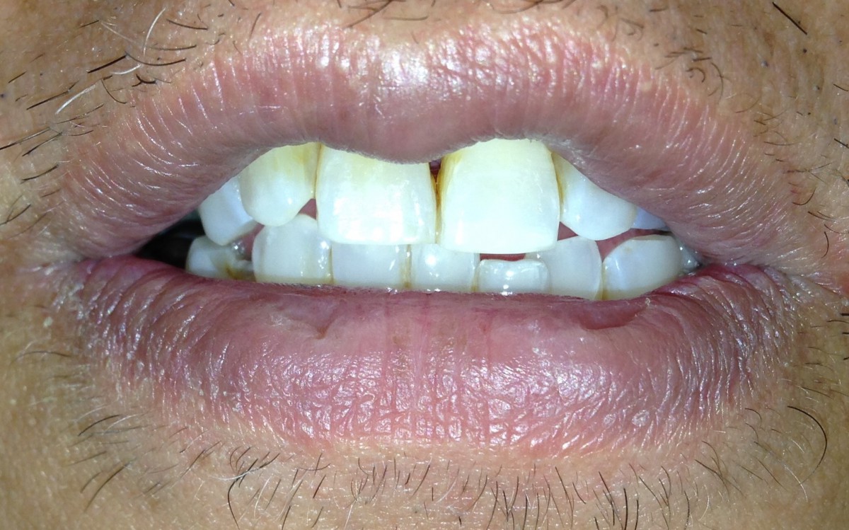
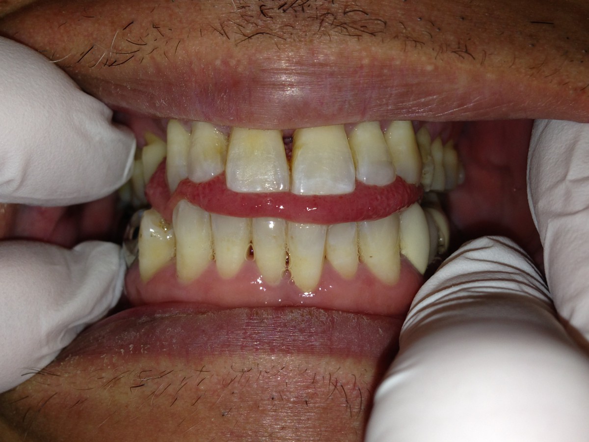
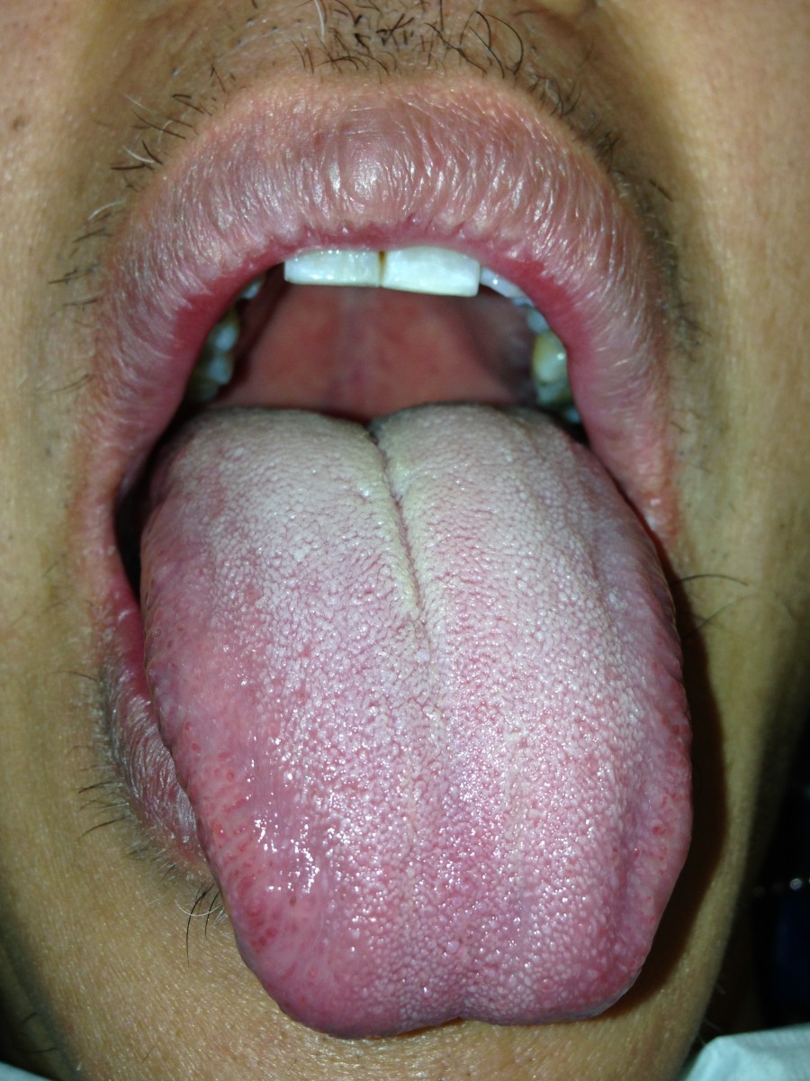
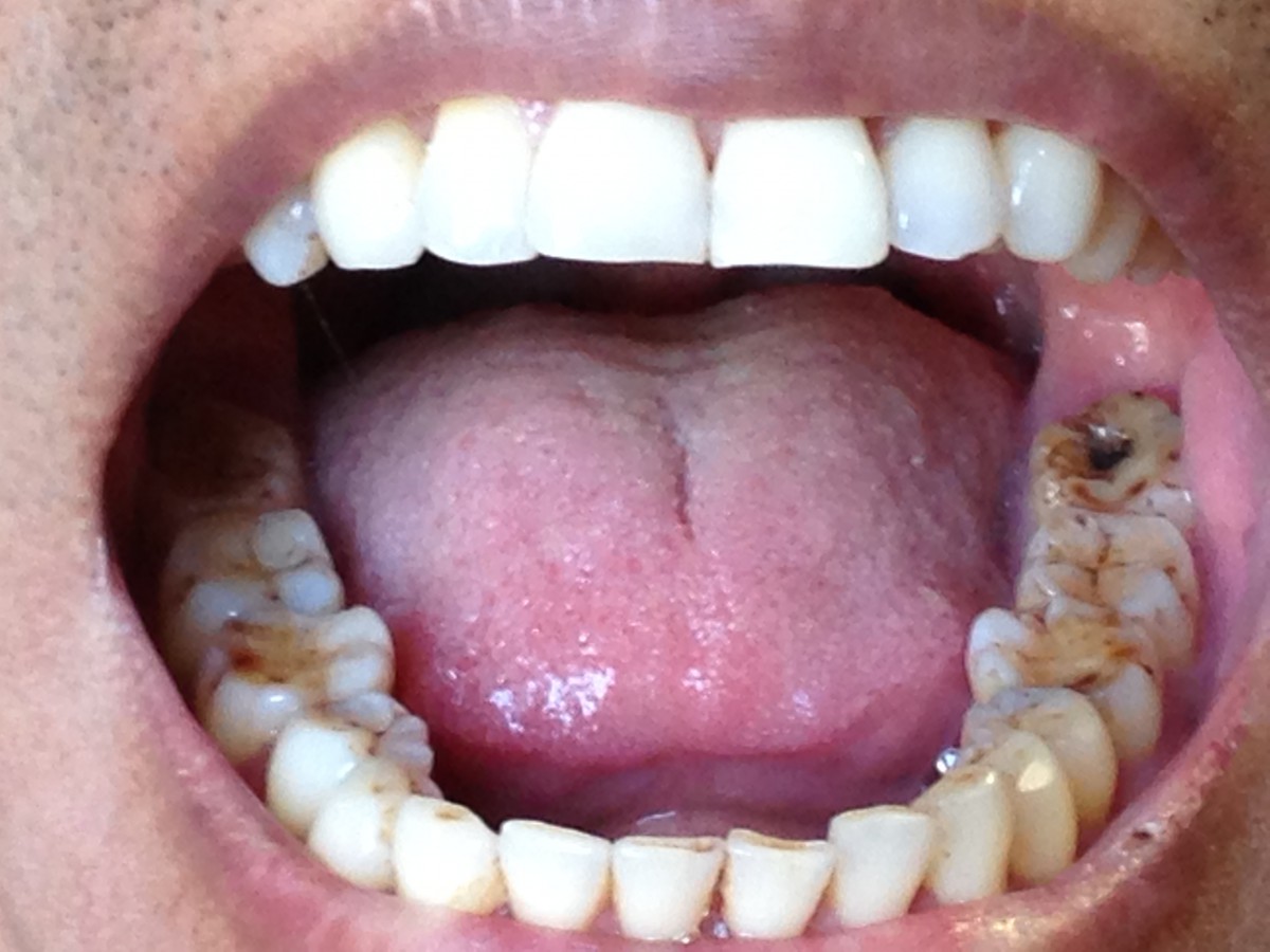
Mallampati score 4
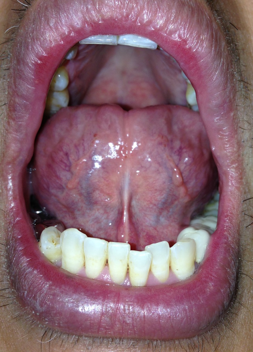
Ankyloglossia
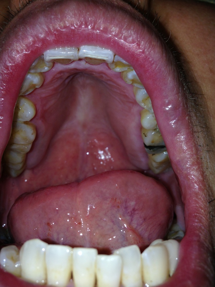
High vaulted palate
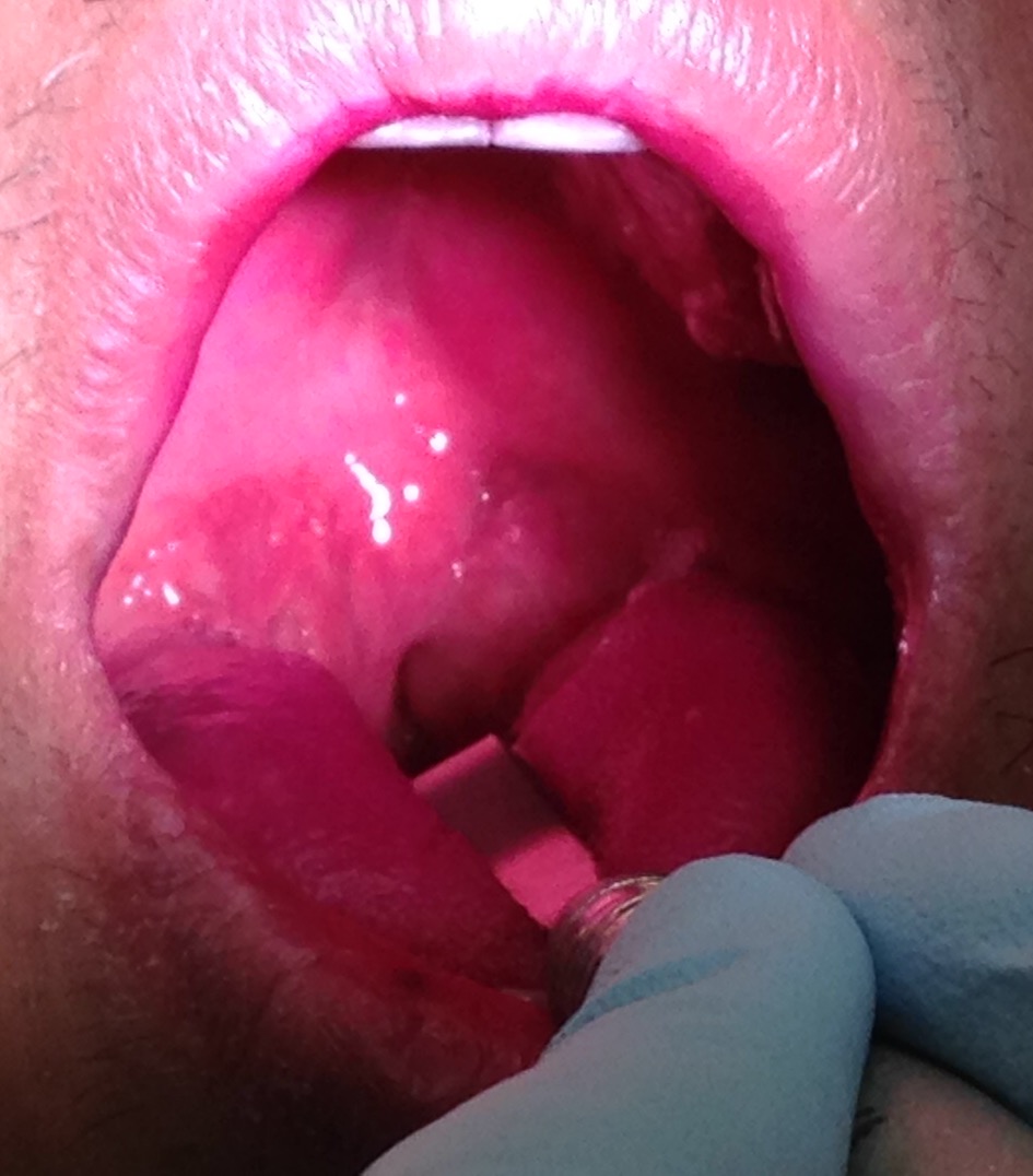
Hypertrophic tonsils
I had a heavy/type II perio case with signs of a sleep breathing disorder due to an orofacial myofunction disorder. The patient was a 45 years old Asian non-smoking, healthy male with seasonal allergies. The intra-oral examination revealed a white coated tongue, along with signs of an orofacial myofunctional disorder. Myofunctional disorders are muscular disorders pertaining to the head and neck. They impact directly and indirectly stability of orthodontic treatment, speech, swallowing and many more. Ankyloglossia was present, along with a forward head posture and a high vaulted palate, indicative of a decreased upper nasal airway space. Hypertrophic tonsils and a Mallampati score 4 were also observed demonstrating signs of a possible airway obstruction. FMS radiograph was taken 5 months ago. The x-ray revealed calculus and bone loss in some posterior teeth. Oral health instructions were given by demonstrating Modified Bass tooth brushing method and flossing. Periodontal scaling and root planning were performed. The patient was referred to an ENT doctor for the evaluation of his enlarged tonsil and signs of a sleeping disorder. Patient was told to follow up with a sleep study in order to help rule out sleep apnea.
Case 2
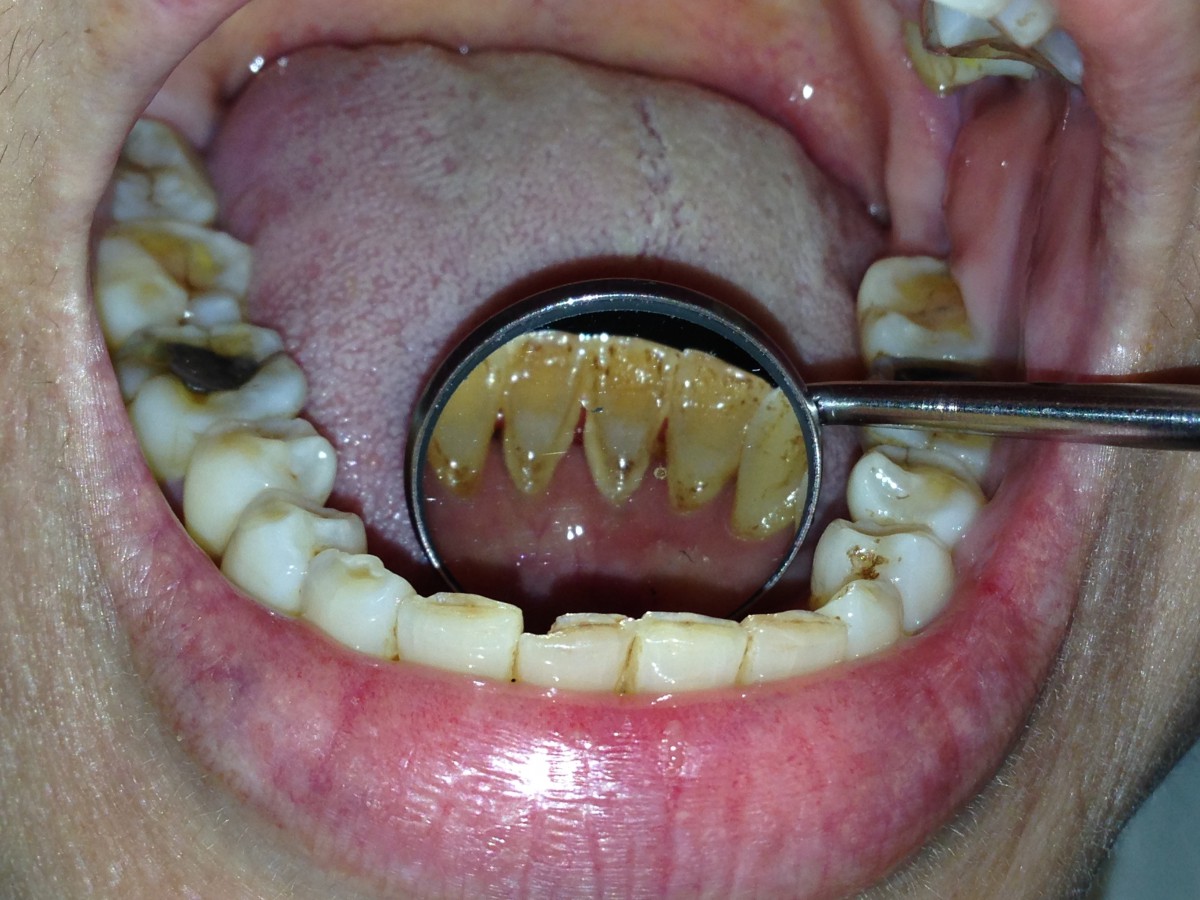
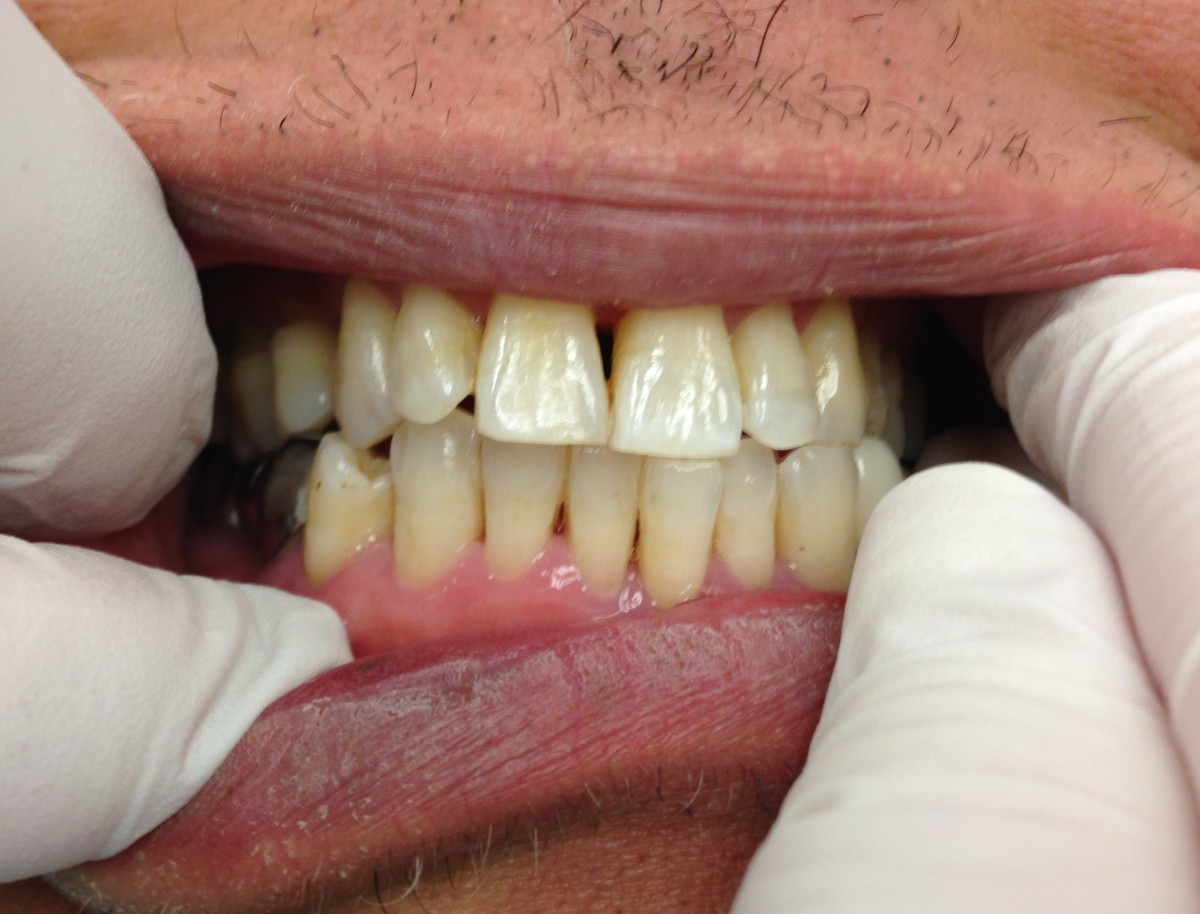
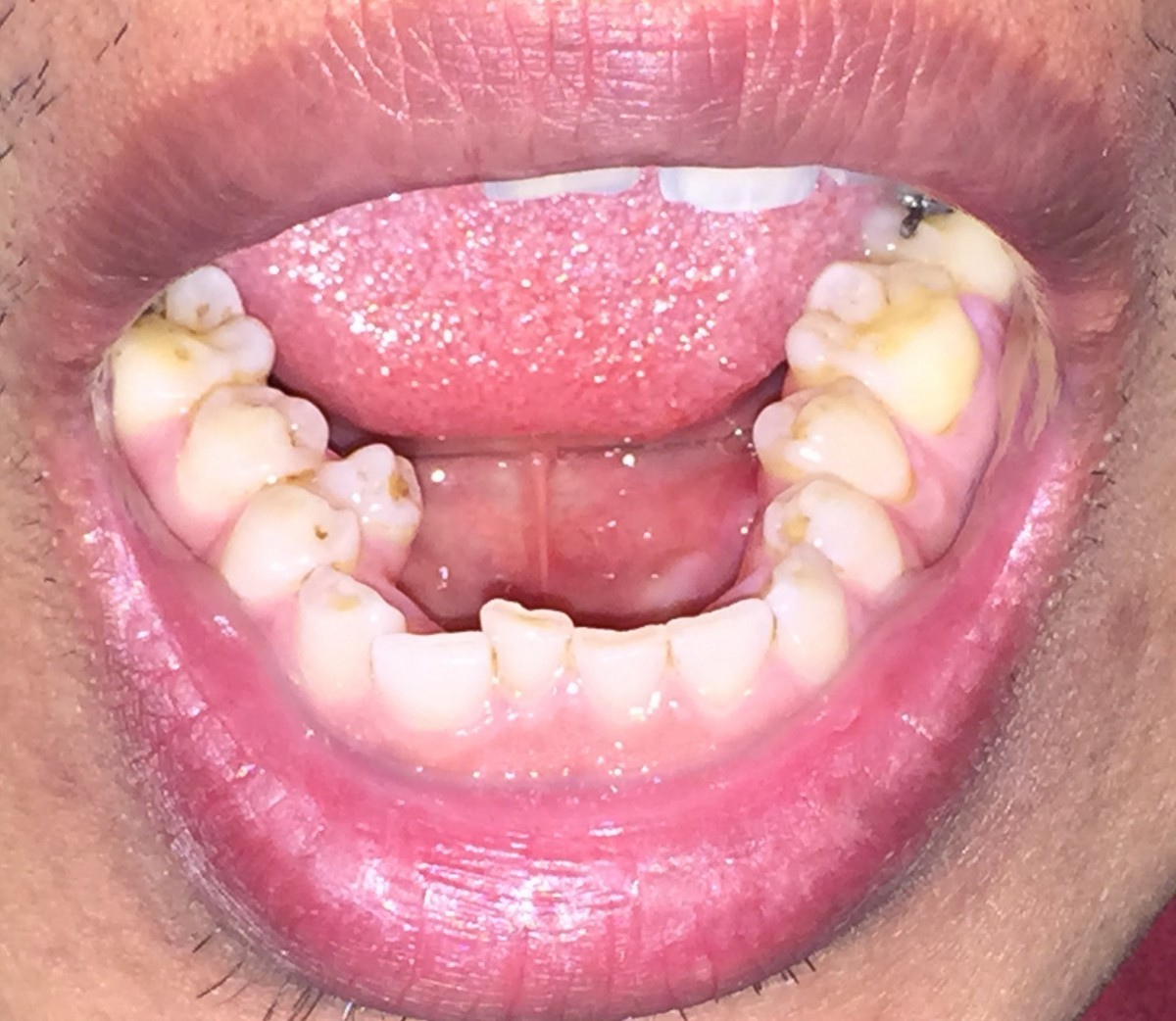
Before teeth cleaning
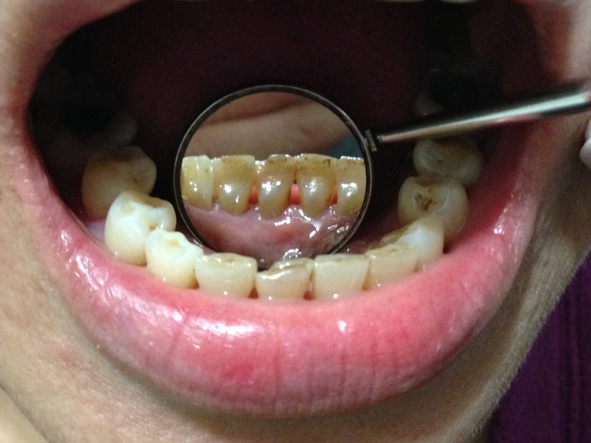
After teeth cleaning
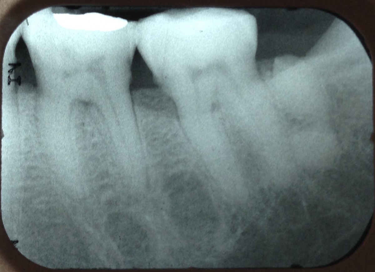
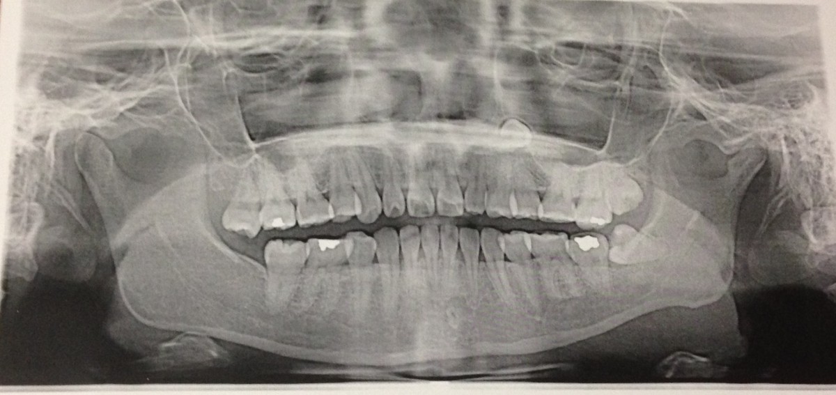
Odontoma
I treated a patient with medium/type II periodontitis. She was a 54 years old Asian healthy female. There was no history of allergy or smoking. FMS radiograph was taken. The x-ray revealed calculus, bone loss and cavity in some posterior teeth and an odontoma around the distal portion of # 18. I provided oral health care instructions, along with dental hygiene prophylaxis.
Case 3
 I treated a heavy/type III perio-case with Arestin placement. The patient was a 45 years old Asian healthy male. No history of allergy or smoking was present. FMS radiograph was taken in 2014. The radiographs revealed calculus and bone loss in some posterior teeth. I provided the patient with oral health instructions, dental hygiene prophylaxis and soft tissue management, along with the placement of Arestin on 5 mm periodontal pockets. Pocket depths decreased by 1 mm. Tissue tone showed improvement. Gingival inflammation and bleeding decreased after 4-5 weeks of treatment. The effectiveness of Arestin therapy was significant.
Case 4
I treated a patient with heavy/ type II periodontitis. The patient was a 27 years old Asian healthy female with history of allergy to dust. Patient stated that 4 teeth were extracted before orthodontic treatment. 4 Bite-wings and panoramic radiograph were taken. Bite-wings revealed calculus on some posterior teeth and caries on occlusal surface of tooth # 17. Panoramic radiograph revealed an impacted canine near roots of tooth # 12 and 13. I provided the patient with oral health care instructions. Periodontal scaling and root planning were performed. Patient was referred to a dentist for evaluation. She was also referred to an oral surgeon for evaluation and treatment of the impacted canine and partially erupted teeth #16 and 17.
Case 5
 I treated a patient with medium/ type I periodontitis. The patient was a 47 years old Asian healthy male. A supernumerary tooth was present near the lingual surfaces of teeth # 28 and 29. It erupted at the age of 37. I provided the patient with oral health care instructions and dental hygiene prophylaxis.

