Just a few case studies of patients varying in age, race and dental conditions. This should give a broad scope into the different scenarios and treatment plans rendered while in clinic
♦♦♦♦♦♦♦♦♦♦ CASE 1 ♦♦♦♦♦♦♦♦♦♦
56 yr old African American male BP=138/78, pulse=69 with history of keloids. Pt came in with pathology on the buccal and lingual of crown #19. Pt explained that an infection occurred after the crown placement 1 year ago but he failed to take appropriate antibiotics and care to dissipate the infection. After appropriate radiographs and assessments, Pt was given a referral to an oral surgeon. At next visit, within a span of two weeks, Pt had tooth extracted. With permission from patient’s surgeon, an SRP was implemented at second visit. Gingiva was generalized moderate edematous erythematous with localized moderate flaccid spongy gingiva around sextant 5. Pt was able to visit weeks later to evaluate gingiva and healing of extraction. Gingiva is now resilient, pink with minimal flaccid spongy gingiva around sextant 5.
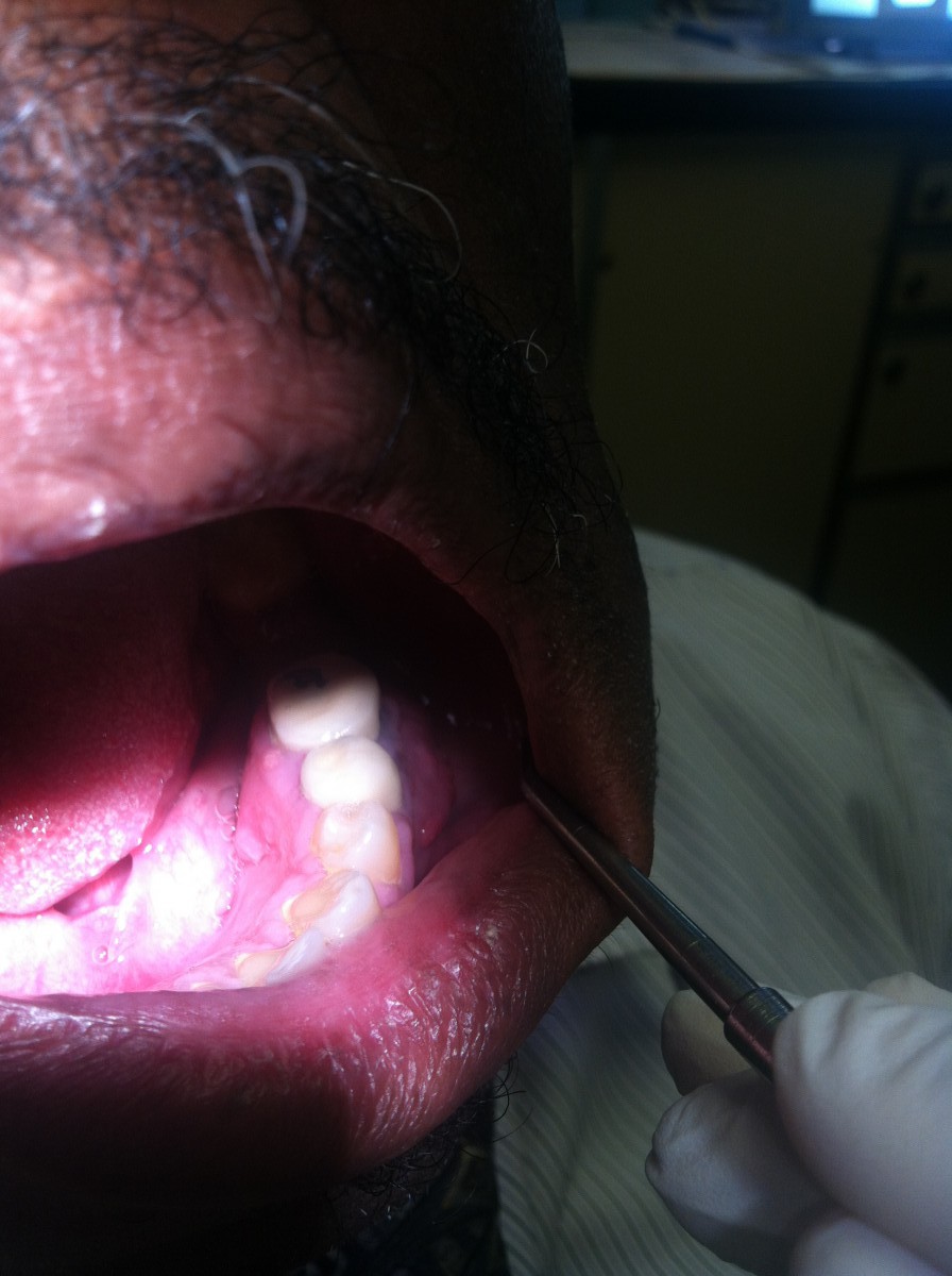
Visit one with pathology; patient given referral
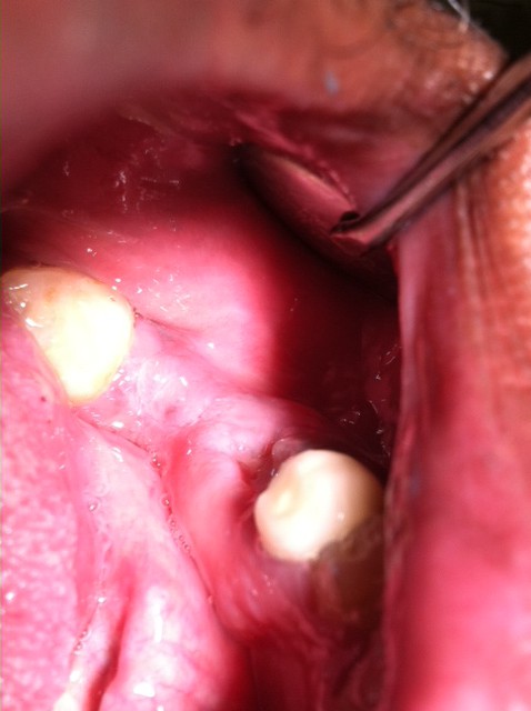
Visit two after extraction at private surgeon office
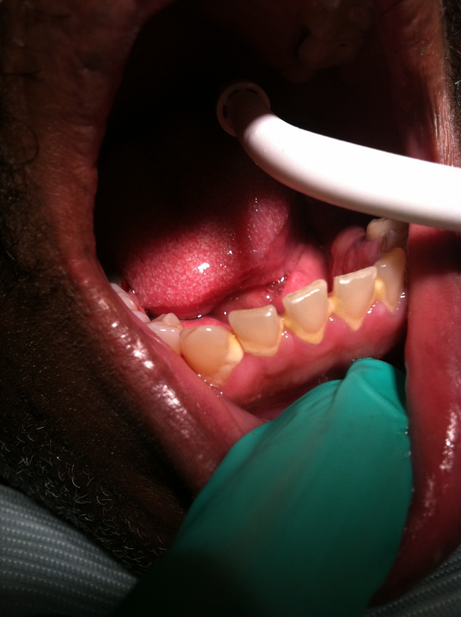
Visit two prior to cleaning
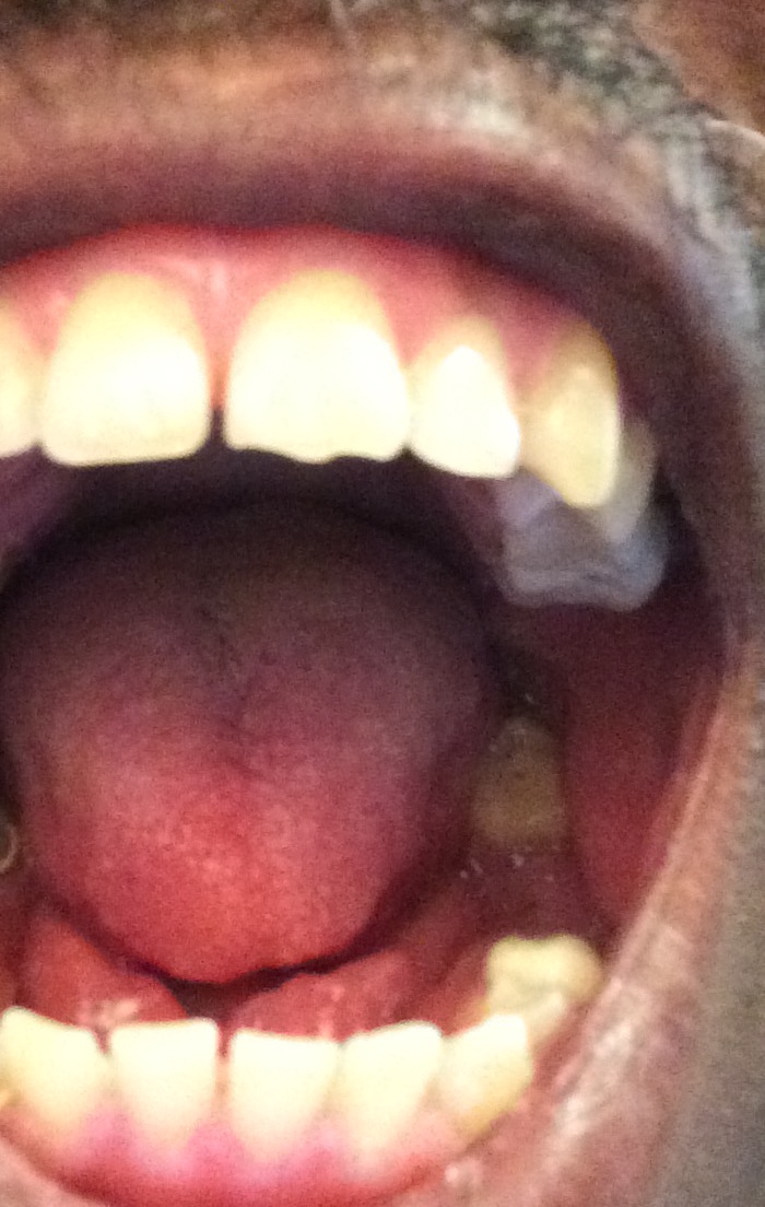
Re-evaluation
♦♦♦♦♦♦♦♦♦♦ CASE 2 ♦♦♦♦♦♦♦♦♦♦
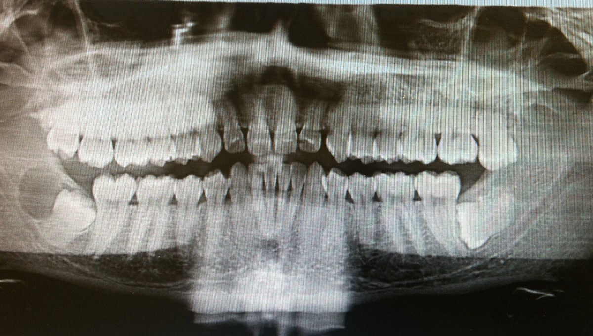
15 yr old female of Indian descent. Pt has mixed dentition however the eruption patterns were out of order and delayed. Pt was given a PANoramic radiograph to visualize and understand which teeth were impacted and why. Pt was treatment planned for a prophylaxis, taught proper hygiene with recommendations, application of fluoride varnish and given a referral.
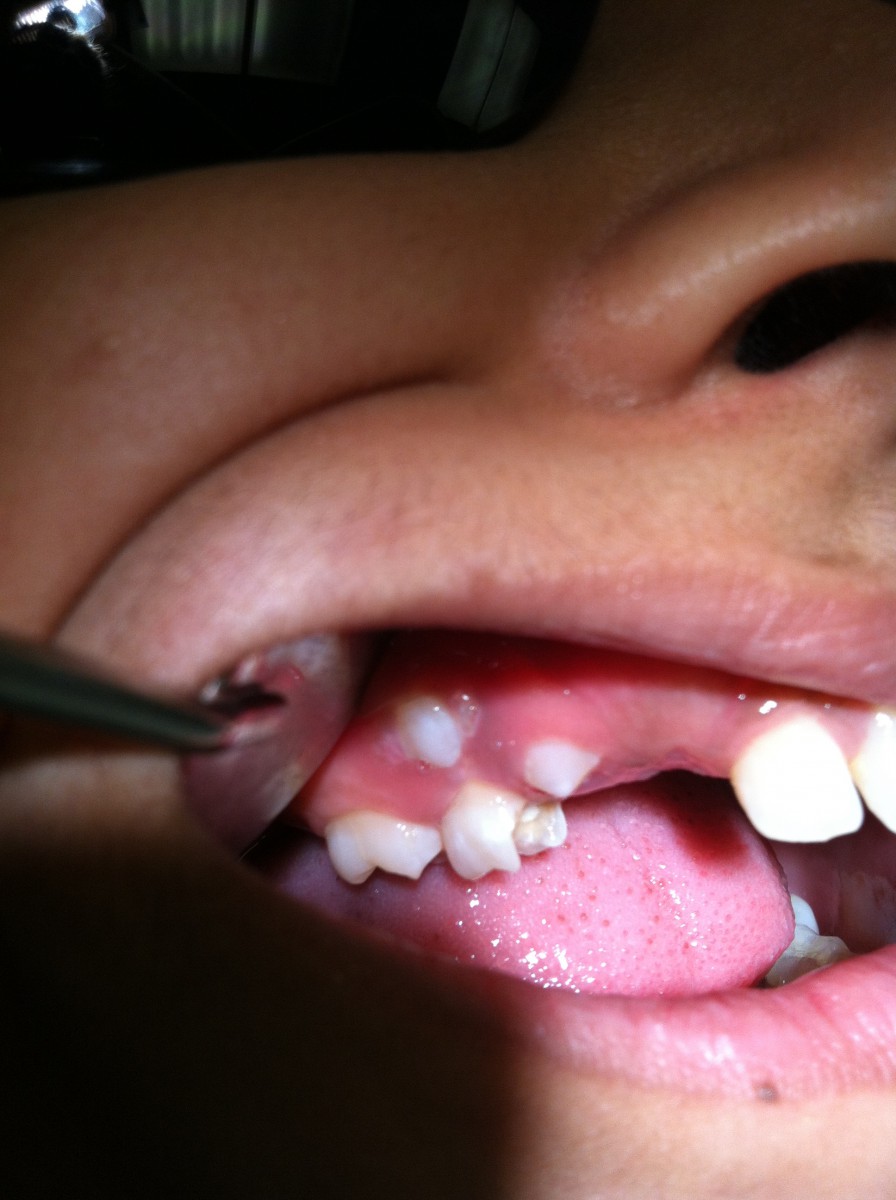
Also notice the missing restoration on MO of deciduous molar
♦♦♦♦♦♦♦♦♦♦ CASE 3 ♦♦♦♦♦♦♦♦♦♦
30yr old male of Caucasian descent. BP=118/78 Pulse=68. No history of allergies, medications or surgeries. Pt was classified as a light, type I periodontal case with localized II around the mandibular molars. Pt required Panoramic radiograph due to unerupted third molars. Upon reviewing radiograph, pt exhibited a pathology around unerupted #32. Pt was given referral for oral surgeon.
♦♦♦♦♦♦♦♦♦♦ CASE 4 ♦♦♦♦♦♦♦♦♦♦
40yr old Hispanic female. BP= 128/76 Pulse=78. Pt classified as heavy with periodontal type II. Pt Caries Risk Assessment determined as High Risk. Took digital bitewings and Panoramic image to assess clinical caries. Pt received hygiene instructions and recommendations, SRP, fluoride varnish and referral for caries.
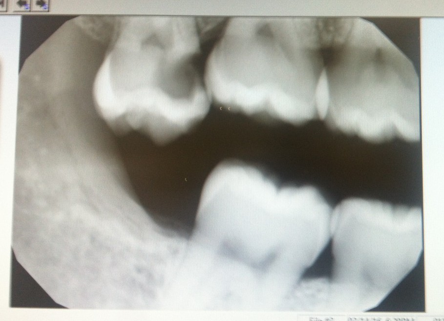
Image of carious lesion. Referral given after completed SRP

