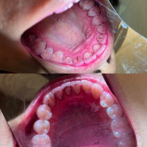
Black-line stain is a highly retentive black or dark brown calculus-like stain that forms along the gingival third near the gingival margin. The stain can be continuous or an interrupted fine line formed by pigmented spots about 1 mm wide. BLS can be seen on facial and lingual surfaces following the contour of the gingival margin onto the proximal spaces. BLS is composed of microorganisms embedded in a ferric sulfide-phosphorus-calcium matrix. These microorganisms are primary gram-positive rods. BLS tends to form over and over again despite regular personal care. There is no definitive etiology but may be from factors such as dietary habits, iron supplements, Actinomyces (bacteria) growth of the black pigmentation. Conflicting data exists about a connection between this type of stain and poor oral hygiene. (Wilkins’ 13th Edition Clinical Practice of the Dental Hygienist)
My clinical experience:
PATIENT PROFILE: Ms. N is a 22-year-old Asian Female. She lives in Brooklyn NY. She came to the dental school clinic for a routine dental cleaning with concerns of black stain on her teeth.
CHIEF COMPLAINT: “I need a cleaning and why is there black stain on my teeth”.
PAST DENTAL HISTORY: Ms. N says she had orthodontic treatment at 10 years old that lasted 1.5 years. She has retainer but does not wear it. Ms. N’s last dental exam was performed on 8/17/2020 which included BW radiographs and a dental cleaning. Ms. N states she has sensitivity sometimes to cold foods on her anterior teeth and she brushes with a manual soft bristled toothbrush 1x a day. Ms. N typically uses Crest brand toothpaste, no oral rinses, and only flosses when needed. Ms. N states that she feels she grinds her teeth at night and sometimes wakes up with a sore jaw.
MEDICAL HISTORY SUMMARY: ASA II (seasonal allergies), Ms. N reports no known drug allergies but does have seasonal allergies with symptoms involving runny nose. Ms. N does not take and medications or OTC vitamins. Last physical exam was on 9/25/2020- MS. N reports good health overall. Ms. N reports 1 alcohol drink per week and non-smoking.
CLINICAL FINDINGS: Extraoral examination: Ms. N has slight bilateral TMJ crepitation that she says is asymptomatic. Intraoral examination: Small nodules felt on buccal mucosa at corners of her mouth. Operculum noted on the distal of #31. Ms. N’s third molars were not clinically present and she states that she never had them extracted. Generalized black line stain was observed on all lingual surfaces as well as buccal and interproximal surfaces. Ms. N’s occlusion was bilateral class I with an overjet of 2 mm and an overbite of 5%. Gingival assessment: Ms. N’s gingiva appeared generalized pink in color with slightly puffy and inflamed interdental papilla on her lower anteriors #22 – #27. Periodontal findings: Pocket depths measured were generalized 2-4 mm with minimal BOP. Localized 5 mm PD was charted on #5ML and #18DL. Calculus detected was localized light sub/supragingival on lower anterior lingual surfaces. Ms. N’s PI score was 2.16 (poor) with heavy marginal biofilm build up on buccal and lingual surfaces. Radiographic Findings: No radiographs were exposed during Ms. N’s appointment.
PLANNING: 2 appointments were scheduled to complete Ms. N’s dental cleaning. Appointment 1 consisted of completion of all patient assessments, OHI recommendation for manual modified bass technique and recommended whitening toothpaste. I advised Ms. N to brush 2x a day (AM/PM). Q1 & Q4 showed heavy extrinsic staining where 90% of stain was initially removed from surfaces using only hand instrumentation due to limited use of the ultrasonic (COVID-19 protocol at the time). These quadrants were polished with pumice and hydrogen peroxide mix. Appointment #2 consisted of review of OHI and I asked Ms. N if she was able to incorporate 2x a day brushing into her routine which she said she was doing. I introduced the c-shaped flossing technique and stressed the importance of daily flossing to remove interproximal plaque build up. Q1 & Q4 were checked for residual calculus which was felt on #27D & #30D. Residual calculus deposits were removed. I continued with the cleaning in Q2 & Q3 with hand instrumentation and was able to use ultrasonic scalers for this appointment. Engine polishing was used again containing pumice and hydrogen peroxide mixture. After the cleaning was completed in Q2 & Q3, ultrasonic scalers were used in Q1 & Q4 to remove additional black line stain. Great results were observed as ~95% of the stain was removed from Ms. N’s dentition. All contacts were flossed and Ms. N was dismissed. Ms. N was instructed to return for her next recare dental cleaning in 4 months where a PAN radiograph should be exposed to check for 3rd molars and fluoride varnish to be administered on both arches.
IMPLEMENTATION: The treatment proceeded as planned and the patient was happy with stain removal results. No skill roadblocks were encountered with dental health education. The patient was able to demonstrate adequate skills using the modified bass method and string floss. Hygiene procedures were accomplished with a moderate form of difficulty due to the heavy extrinsic stains. Photographs were taken and permission was obtained to use them.
Before and After-Buccal and Proximal Surfaces
Before and after-Maxillary Lingual Surfaces Clinical BLS-Maxillary and Mandibular Lingual Surfaces


