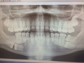
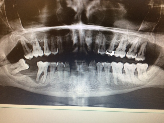
Photo #1
Shows a panoramic film that I had exposed on my patient. This patient had mobility on all molars, with heavy calculus, and severe bleeding. A referral was given for the removal of all molars. Scaling and root planing was part of my treatment plan, with the use of local anesthesia. When the patient came back for the second visit, #17 had been extracted, and the previously scaled area already shown great improvement. My patient proved to me that my oral hygiene instructions helped to reduce inflammation and bleeding. This patient has radiographically seen calculus that was a challenge to remove, but I was successful in removing it all.