Demographics:
-30 year old
– Hispanic
-Male
Assessment:
-Blood Pressure: 125/78, Pulse 76.
-ASA II: Patient takes Metformin- 500 mg for management of Type II Diabetes. Patient’s last A1C number was 6.5. Patient reported that he used to smoke 1 pack a day for 3 years, but has not smoked for 6 months. Patient has no known allergies, no recent hospitalizations, and doesn’t report any other systemic conditions.
-EO/IO: Asymptomatic bilateral clicking of the TMJ. Bilateral Linea Alba.
-Occlusion: Class I occlusion with 40% overbite and 5mm overjet.
-Home care: Patient reports brushing once a day in the morning, doesn’t floss, and uses an antiseptic mouth rinse 2-3 times per day.
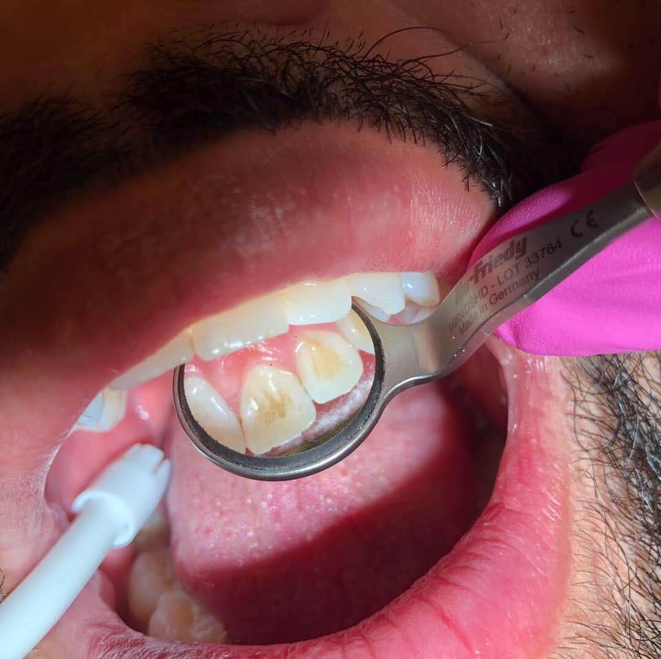
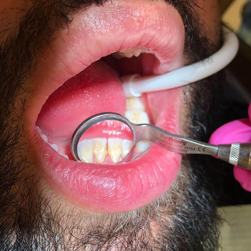
-Deposits: Generalized moderate subgingival calculus, predominantly detected in interproximal areas. Localized light to moderate supragingival deposits on lower anteriors with generalized mild staining. Localized light-moderate staining on lingual surfaces of anterior teeth.
-Plaque: Patient had a plaque score of 1.0 (Fair).
– Generalized pink, spongy, pointed gingiva that fills the interdental space.
– Type I active gingivitis, without radiographic evidence, due to generalized 3-4mm probe depths, moderate bleeding upon probing, and localized recession on lower anterior teeth. No furcation activity or mobility observed.
Planning
I was able to formulate the treatment plan and obtain informed consent in the first visit. The tx plan was to scale two quadrants per visit using local anesthesia for patient comfort.
Treatment plan was as follows:
V1: Expose radiographs. Plaque index. OHI: Teach patient proper flossing technique. Scale UR and LR quadrants to completion with the ultrasonic and hand scalers using local anesthesia.
V2: Plaque index: OHI: Review flossing technique. Teach patient the Modified Stillman toothbrushing method. Re-evaluate UR/LR quadrants. Scale UL/LL quadrants to completion using local anesthesia, ultrasonic and hand scalers.
Implementation:
V1: Four bitewing radiographs and one panoramic radiograph exposed to assess for calculus, caries, bone loss, and pathology.
Plaque index was performed. This patient’s plaque score was 1.0 fair. Majority of plaque build up was observed on the on the interproximal surfaces of all teeth, and on the cervical third of the lingual surfaces of posterior teeth.
OHI: Patient was taught the proper flossing method. Patient was able to correctly re-demonstrate what he was taught. He explained that he was never taught how to properly floss before. Patient seemed motivated to continue flossing. I instructed this patient to begin flossing at least every other day.
Polished the LR quadrant with course prophylaxis paste to remove stains.
Scaled UR/LR quadrants to completion using the ultrasonic and hand scalers. Oraqix (2.5% Lidocaine, 2.5% Prilocaine) used for patient comfort. Patient tolerated procedure well.
V2: New plaque index was performed. This patient’s plaque score was remained 1.0 (Fair) due to plaque build up on the cervical third of the lingual surfaces of posterior teeth.
OHI: Patient was taught the Modified Stillman’s toothbrushing method. Patient did not seem motivated to continue this method, so I introduced the Sonicare electric tooth as an alternative. Patient was able to purchase the toothbrush at this visit. I demonstrated the proper use and care of this toothbrush.
Re-evaluated UR/LR for residual calculus. Residual calculus was observed on #25-L and #27-DL. I scaled those surfaces to completion.
Polished the linguals of the anterior teeth with coarse paste to remove stains.
I scaled the UL/LL quadrants to completion using the ultrasonic and hand scalers. Polished entire dentition with fine paste. 5% Fl varnish treatment was applied. Patient was placed on a 4 month recare regimen.
Evaluation:
-The patient was compliant with OHI recommendations. Although PI showed no improvement, a 50% decrease in bleeding was observed. Gingival tissue appeared pink, pointy, and resilient.