
25 years old, white male, Medium Type I

A 25-year old male presented for an initial visit. Reviewed medical history – WNL. Patient is non smoker, non drinker, no medications are taken. ASA I. Last dental visit as well as dental cleaning were 6 years ago in March 2013. FMS were exposed at that time. Patient reported to use medium filaments tooth brush, to floss three times a week and to use the back of his tooth brush as a tongue cleaner. Patient said he does not use any oral rinses.
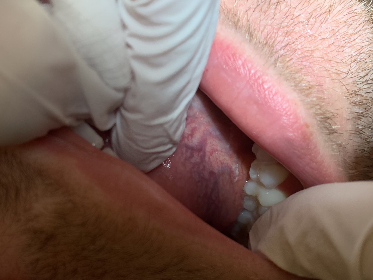
Extra-oral examination: WNL. During an Intaoral exam a small white leukoplakik lesion was noticed on the right lateral service of the tongue, close to the floor of the mouth. The lesion was asymptomatic and could not be rubbed off. Patient reported to see it before. Patient emphasized that the lesion becomes bigger with consumption of acidic food. Patient was informed that Leukoplakia is a broad term that defines lesions that appear as white patches found in the inner lining of the mouth. Leukoplakia can be caused by multiple reasons, therefore its treatment varies. Most of the time, leukoplakias are painless, but it’s extremely important to identify the source of the condition, as some leukoplakias indicate more serious problems, like autoimmune disorders, and can even develop into oral cancer.
After completion of the treatment, patient was referred to the oral pathologist for the further evaluation of the lesion.
As per today, patient reported that he did not visit an oral pathologist yet.

