
There are several clinical cases I encountered that enhanced my knowledge of patient care. One case consisted of a patient who had active periodontal disease. Probing depths ranged from 3-9mm. After completion of my assessments on this patient, I decided it would be a good idea to place Arestin/minocycline HCL 1mg in periodontal pockets measuring 5-7mm to kill the red complex bacteria in that area that is advancing the disease. Red complex bacteria causes progression of periodontal disease if undisturbed.
Case 1: Arestin

Here is my Arestin patient prior to treatment.
S.L. 53-year-old male M/III
Date of first service: October 5th, 2017
ASSESSMENTS:
Medical History: Patient ‘s medical history is WNL. ASA I. Patient experiences seasonal allergies. Patient has had gingival flap surgery on maxillary anteriors. No premedication necessary. Patient’s blood pressure measured 119/69. No systemic conditions. No Medications.
Oral pathology. Crepitus of the TMJ was present on the patient’s right side during lateral excursions. Patient has a scar on the dorsal surface of his tongue. Patient stated that the scar has been there since childhood due to physical injury. No referral given for pathology.
Dentition: Generalized attriton is present. Patient is missing tooth #1, 16, and 17. Patient has Class I occlusion with a cross bite on his right side. Possibly caries active. Possible caries present on the occlusal of #32 and cervical third of #21. Patient is high risk for caries due to; teeth being lost due to caries, consumption of beverages containing high sugar concentrations, presence of possible caries, family history of caries and several dental restorations.
Periodontal: Gingiva was generally fibrotic possibly due to past history of smoking. Generalized inflammation was present around the gingival margin and interdental papilla. Posteriorly the tissue was spongy and blunted, anteriorly the tissue was more firm and knife edged. Minimal to moderate BUP. Perio Type III. Probing depths ranged from 3-9mm. Generalized recession was present. Patient was approved for an FMS. Patient is periodontal disease active. This patient qualified for Arestin. He had several pocket depths ranging from 5mm to 7mm. Most of these probing depths were located in molar and premolar region.
Oral hygiene: Patient had medium amounts of calculus deposits. In order to assess his oral hygiene I first conducted a plaque score. His plaque score was .7. Most plaque accumulation was interdentally. I demonstrated proper flossing to the patient in order to disrupt the plaque biofilm causing his deep pocket depths. I allowed him to demonstrate flossing on his own and he was having trouble reaching his hands to the back of his mouth. I recommended using a floss holder in order for him to have better access to the posterior teeth.
Radiographs: Exposed FMS. FMS revealed possible recurrent decay on #30-D and horizontal bone loss anteriorly and posteriorly.
Time: The time interval between his last dental visit and the current visit was not appropriate. A seven-month time span is too long for a periodontal disease active patient. This patient should be placed on a 3-month recare schedule in order to disrupt any bacteria contributing to the progression of his periodontal disease.
Dental Hygiene Diagnosis: Patient had active periodontal disease, was possibly caries active and experiences localized dentin hypersensitivity on facial of exposed roots of mandibular canines and premolars.
Treatment Management/ Plan/Implementation:
Treatment plan: Provide oral hygiene instructions at each visit. Expose FMS. Scale all quadrants using hand instruments and ultrasonic. Referral to patient for oral conditions. Fine engine polish. Administer 5% NaFl Varnish.
Treatment rendered at visit one included; oral hygiene instruction and scaling of all quadrants using hand instruments and ultrasonic. Referral was given to patient for evaluation of possible caries and periodontal evaluation. Fine Polished. Administered 5% NaFl Varnish.
Treatment rendered at visit two included Arestin placement on #2MB-7mm, #2MF-7mm, #14DL-6mm, #15ML-6mm, #15MF-5mm, #15ML-6mm, #19DL-5mm, #32MF-7mm, #30DF-6mm. Postoperative instructions were given to patient.
Treatment Rendered at visit three included re-evaluation of areas treated with Arestin. Probing depths of the treated areas measured: #2MB-5mm, #2ML-6mm. #32MF-4mm #30DF-5mm #15ML-5mm &MF-3mm #19DL-4mm #14DL-4mm.
Evaluation: Patient responded well to treatment interventions done at each visit. His tissue responded well to the treatment. Inflammation and bleeding had subsided. Localized inflammation was present on the buccal aspect of #7 around the gingival margin and interdental papillae. There was minimal pocket reduction on premolars (1mm reduction). The patient was happy with how thorough the visit was. He was motivated to improve his oral hygiene routine and was excited to complete treatment. He asked many questions about using a power toothbrush and he also explained that he has been using both the waterpik and manual floss to clean between his teeth. He stated that he feels like gets a better clean in certain areas with the manual floss, but is able to access harder to reach areas using the waterpik. I advised patient to switch over to an electric toothbrush in order to improve his total removal of plaque biofilm.
Patient was placed on a 3 month re-care schedule.
Documentation: The assessments about this patient and the treatment that I provided along with the patients response to treatment and treatment outcomes are clearly written in the patient’s chart for future reference of other dental professionals within the NYCCT clinic and for legal purposes as well.
Case 2: Medically Compromised patient.
D.F. 47 year old male. M/II
Visit Date: 5/9/2018
Assessments:
Medical History: Patient is ASA II due to hypertension and heart murmur. Patient is taking Metoprolol 50 mg 1x a day to manage hypertension, and baby aspirin 80mg daily to combat chronic knee pain. Patient had catheter ablation done to correct tachycardia. Patient has seasonal allergies. Patient’s blood pressure measured 144/91. Pulse was 61.
Oral Pathology: Patient presented with a 1x1cm hemorrhagic exophytic mass consistent with soft tissue hemangioma, patient consented to have photo taken. He stated has seen his dermatologist for the hemangioma, but has not set up an appointment to have it drained. Referral was given to patient for evaluation of hemangioma by the oral surgeon.
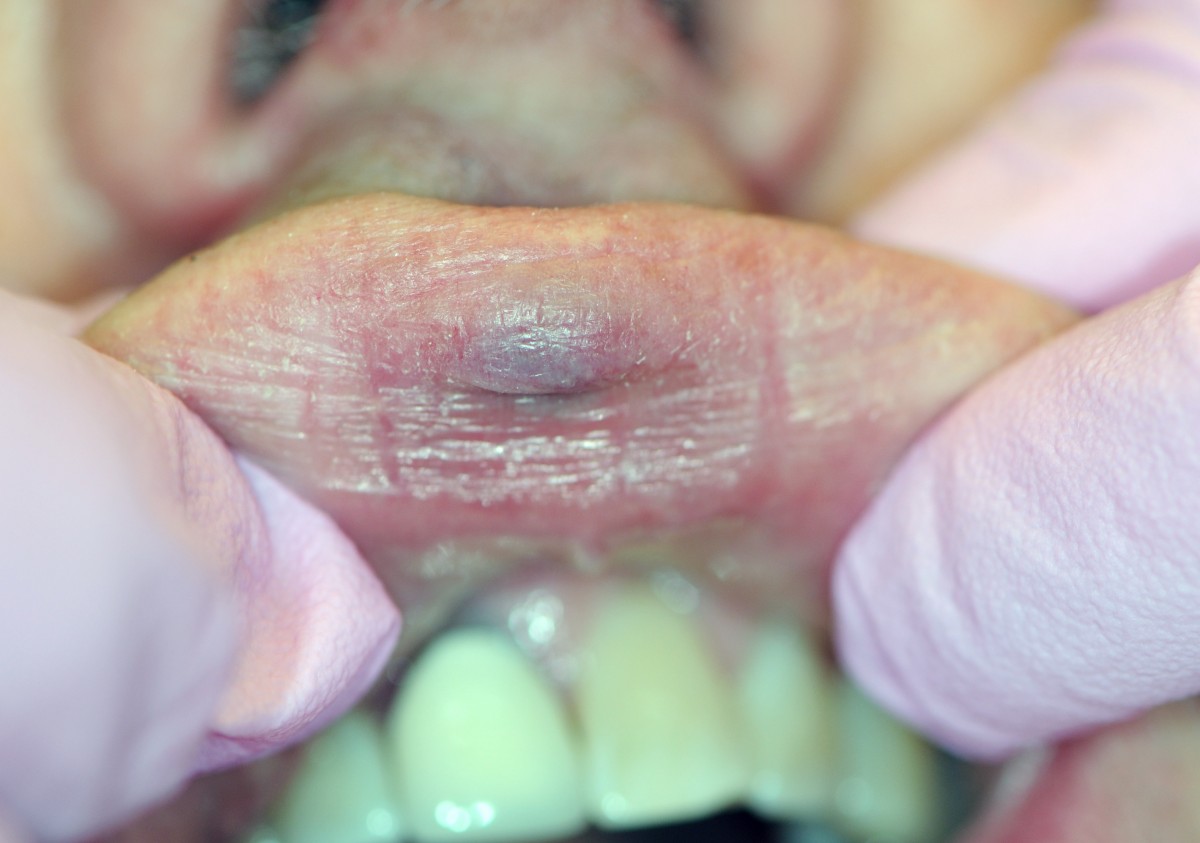
This patient’s Hemangioma.
Dentition: Patient presented with two amalgam restorations present on the occlusal of #3 and #14 along with a porcelain fused to metal crown on #8. Class I occlusion. Generalized mild attrition. Wear facets on occlusal and incisal surface did not signal the need for a mouth guard.
Periodontal: Patient’s gingiva was generally pink and firm with localized inflammation around the gingival margin and interdental papillae of all lingual surfaces. Minimal to moderate bleeding upon probing. Generalized recession present possibly to due toothbrush abrasion. Advised patient to obtain a pressure sensing electric toothbrush to minimize wear of tooth surface and periodontal structure.Probing depths ranged from 3-4mm. Patient is periodontal type II due to loss of attachment when combing 4mm readings and 1-3mm recessions.
Oral Hygiene: Patient stated he does not floss regularly and brushes twice a day with a manual tooth brush and a fluoridated toothpaste. He stated he rinses with a non-alcohol mouth wash. Patient presented with medium amounts of subgingival calculus deposits. I advised him to brush 2x a day with an electric toothbrush that has a pressure sensor as mentioned earlier. I also advised him to floss at least once a day and to try to take “baby steps” by only flossing a couple of days a week and adding a new day each week until he reaches a full 7 days. I also advised him to use anti-gingivitis oral health products (rinses and toothpastes) to combat his mild gingivitis. He has not been to the dentist in 2 years so I advised him to return every six months to reduce gingival inflammation and prevent disease. Supra gingival staining was present on mandibular anteriors and maxillary left posteriors possibly due to tea drinking.
Radiographs: No radiographs were taken at this time. FMS was exposed two years ago and there was no clinical evidence signaling significant changes in his oral health. No clinical evidence suggested the need for radiographs.
Time: The time interval between his last dental visit and his current visit was not appropriate. A two year time span is too long of a time without any prophylactic dental treatment. This patient should be placed on a 6-month recare schedule in order to disrupt any bacteria contributing to the progression of gingivitis into periodontal disease.
Dental Hygiene Diagnosis: Gingivitis.
Treatment Management/ Plan/Implementation:
Treatment Plan: Oral hygiene instruction using manual floss. Calculate plaque index. Scale all four quadrants using hand instruments and ultrasonic. Air polish all quadrants to remove supra gingival staining. Apply 5% NaFl Varnish.
Treatment Rendered: Oral hygiene instruction using manual floss. Plaque index measured .6. Scaled all four quadrants using hand instruments and ultrasonic. Air polished all quadrants to remove supra gingival staining. Referral given to patient for evaluation of possible hemangioma. Applied 5% NaFl Varnish.
Evaluation: Patient responded well to treatment. Patient enjoyed the experience and stated that he regrets not coming in within the last two years to be evaluated and have appropriate treatment. Patient comprehended all home care recommendations and asked for a list of all home care changes I advised him to make. Patient’s medical conditions did not impact the treatment plan.
Patient was placed on a 6 month re-care schedule.
Documentation: The assessments about this patient and the treatment that I provided along with the patients response to treatment and treatment outcomes are clearly written in the patient’s chart for future reference of other dental professionals within the NYCCT clinic and for legal purposes as well.

