Case 1
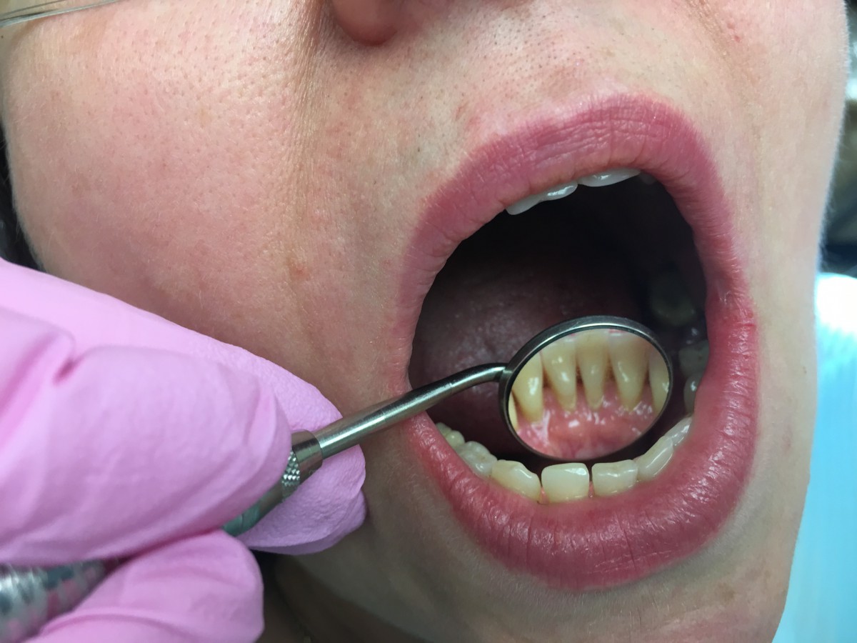
46 year old caucasian female. Med history WNL. Extensive root caries, Type II embrasures with generalized recession. SRP and OHI performed. Referral given to dentist for root caries evaluation and periodontist for complete periodontal exam.
Patient had extensive caries due to root surface exposure. Most surfaces had arrested decay, however some were soft. There were heavy subgingival and supragingival generalized deposits. Patient was interested in taking care of her caries and will follow up with her dentist.


Case 2
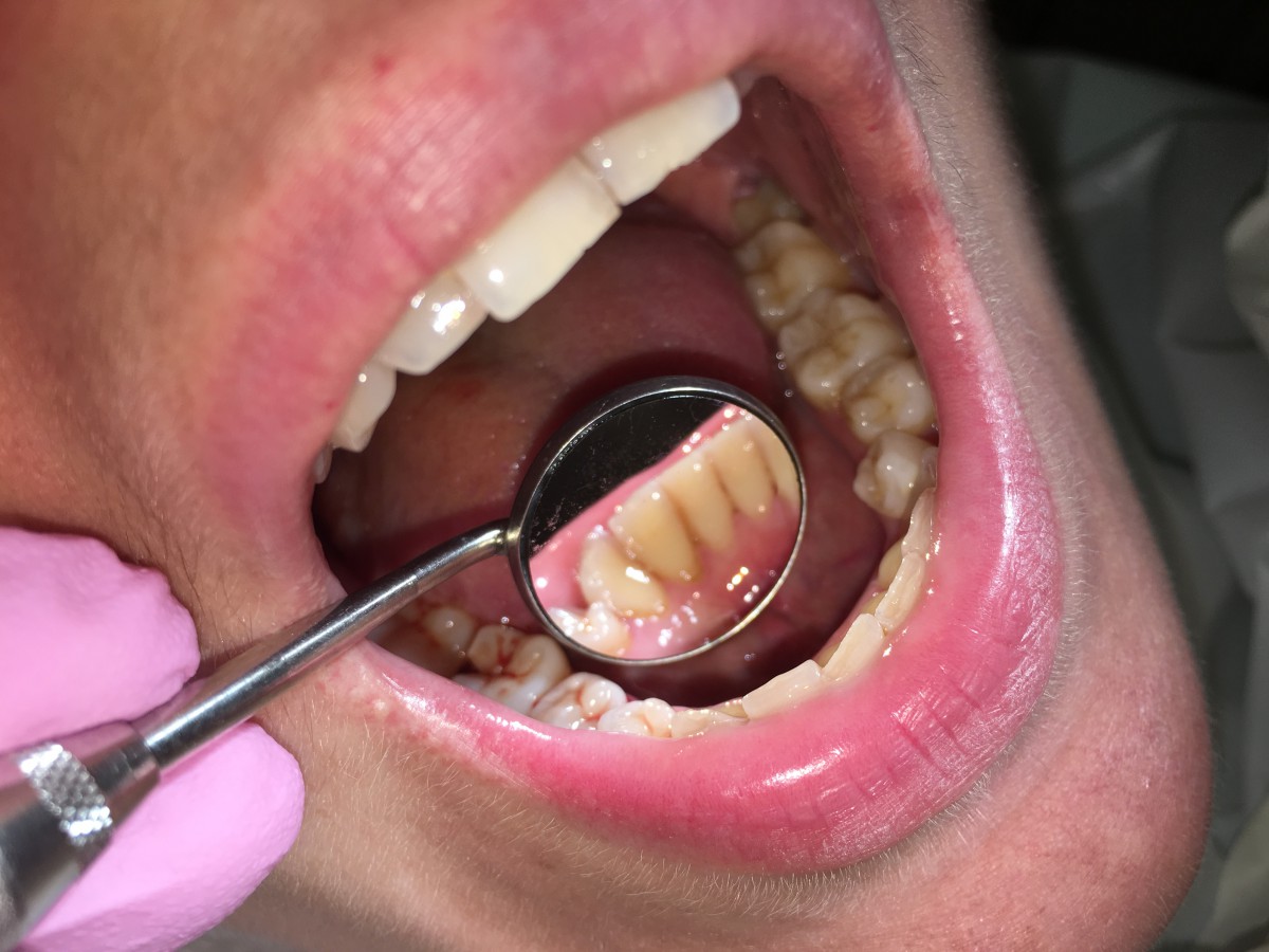
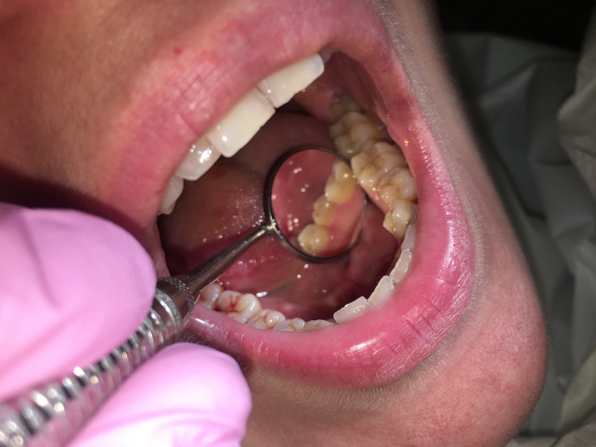
33 year old caucasian female. Medical history WNL. Generalized gingivitis and heavy subgingival deposits. Performed SRP and OHI. Patient was compliant and interested in improving oral hygiene.


Case 3
Here I would like to share one of my journal written on a patient I had in my freshman year in clinic:
Demographics
Patient initials L.K. age 44, heavy/ type II.
Assessment:
- Patient history. Patient was given Amoxicillin 500mg once before treatment when bone graft procedure was done. Vital signs were within normal limits, patient usually has low blood pressure 98/67 and pulse was 84.
- Non-smoker.
c. No premedication required. - Patient has allergies to lidocaine, penicillin citruses, silver, seasonal allergy, chlorine, shrimps.
- No medicines are currently being taken.
Oral Pathology:
- Extra Oral exam significant finding: soft palpable node on left anterior chain, TMJ clicks right side.
Intra oral findings: bilateral posterior linea alba and palatine torus, ranula on the floor of the mouth left side, on sublingual caruncle (confirmed to be WNL varicosity of the floor of the mouth on last visit).
Dentition:
- Class of Occlusion I. Overjet 3mm. Overbite 50%.
- Patient has extracted #3,12,14,16,30. Abfraction on 13F and 5F. Attrition on 6-11 and 20-29. Patient has a diastema between #8 and #9.
- Caries lesions found on #1 Occlusal and #4 Occlusal.
Periodontal:
- Patient has minimal bleeding on probing, minimal inflammation, Type II Slight Periodontitis. Probing depths range 2-5mm, with bigger numbers present in the molar region. Recession on #22F – 2mm, 29DB – 4mm, 31MB – 4mm. Recession on #22F seems due to chewing, biting and other normal forces. Recession on 29DB and 31MB is due to absence of #30 and destruction of interdental papillae between these two teeth.
- Gingiva is pink in color, resilient, intact, inflammation is minimal, tissue is firm, fills in interdental spaces, is pyramidal and pointy.
Oral Hygiene
a. Initial Plaque Score was 0.5. Revisit plaque scores were 0.5 and 0.5.
- Supragingival calculus was found on Mandibular Anterior Lingual and premolar teeth #20L-28L.
Generalized subgingival calculus was found on Mandibular Anterior Lingual surfaces and most interproximal posterior teeth majority on the right side.
- In session one showed patient Modified Sulcular Bass Technique. In session two flossing was shown with Reach Floss Holder because patient reported difficulty flossing posterior areas. In session three reviewed tooth brushing method and flossing with Floss Holder again.
Radiographs:
Patient had a PA taken on January 2016 of #12. This PA was not available at the time of treatment. Four digital bitewings were exposed during Radiology Laboratory Class after patient has been completed on 3/18/2016. Slight horizontal bone loss was found in molar areas, which supports Type II periodontal classification. Visible interproximal calculus was found on the BWS. No interproximal decay was found.
Treatment Management:
- In the first visit 2/8/16, assessments were completed up to periodontal charting. Medical history was checked, vital signs taken, extra-oral and intra-oral examination completed, dental and periodontal charting probing completed. Dr. Brown identified the soft palpable node on left anterior chain within normal limits.
Revisit 2/22/16. Periodontal Type II assigned after periodontal charting was checked. Heavy case assigned after calculus detection completed. Treatment plan composed of four visits. Patient signed treatment plan. Patient was disclosed, plaque score of 0.5 was recorded. Oral hygiene instructions were given, showed modified sulcular bass technique. Hand scaled #27-32.
Revisit 3/7/16. Extra oral and intra oral re-assessment of previous findings completed. Redness on left side of the floor of the mouth was present. Treated area evaluation revealed gingiva is pink in color, resilient, pyramidal and does not show signs of inflammation. New plaque score recorded 0.5 Oral hygiene instructions reviewed, showed patient flossing with Reach Floss Holder because patient had difficulty reaching posterior areas with regular floss. Hand scaled Upper Right quadrant and Upper Left quadrant, and completed hand scaling Lower Right quadrant. Patient was sensitive on #2, 13F, 24, 25. No anesthesia given as patient is allergic to lidocaine and penicillin. Patient’s first reaction to lidocaine was in 1995 during general anesthesia given to her during the labor of her second child. Symptoms of palpitation and tremor were evident and the doctors noted this in her chart. Second reaction was during dental treatment in 1997. Patient had a molar extraction at that time. The dentist warned her to tell every doctor that she has allergy to lidocaine ever since.
Revisit 3/14/16. Extra oral and intra oral re-evaluation completed. Intra-oral findings revealed redness of floor of the mouth decreased. Right side tonsil was red. Gingiva of #13F was noticed to be inflamed during tissue evaluation. Other gingival tissue showed no signs of inflammation. Patient reports using advised tooth brushing method and flossing more frequently. New plaque score was 0.5. Hand scaled LL. Polished four quadrants. Applied 5% Sodium Fluoride Varnish with Xylitol (Vella Spearmint). Recall length given: 4 months due to heavy case type and rapid deposit formation.
- Patient is allergic to lidocaine, no anesthesia could be given to minimize sensitivity. Advised patient to use Sensodyne toothpaste.
- Recommended Floss Holder for patient for easier access to posterior areas. Patient had difficulty reaching posterior teeth with regular floss. Patient’s response was positive; patient liked using the floss holder but had some difficulty learning at first. Reviewed use of Floss holder in subsequent visits with patient.
- No referral to DDS or MD was given. Patient was contacted after treatment was finished and was advised to see a dentist to treat active caries lesions on #1,4 and possible abfractions found on 5F and 13F.
- Patient was very responsive to treatment and recommendations given. I would not have changed any methods or treatment plan introduced. In my opinion, this treatment plan gave the patient enough time to practice given recommendations, bring back feedback and critique, and enough time between tissue evaluation of treated areas.
Evaluation:
- Patient was motivated and compliant. When the patient did not understand what was being taught, all questions and concerns were answered.
- The patient was more motivated with each treatment progress. Patient reported that teeth were feeling much better and the patient was more motivated to floss because she could feel the food remained between teeth after eating.
- From initial visit patient’s gingiva did not show signs of inflammation; tissue was resilient and pink in color, filled spaces and pyramidal. Through out the treatment the scaled areas did not show signs of inflammation upon revisit evaluation. However, bleeding and sensitivity was present during scaling. Gingiva on tooth #13 Facial surface was inflamed on revisit evaluation; there was no bleeding on probing however the tissue was soft. Other areas presented with healthy pink color and well fitted around the tooth.
- On the last visit, the ranula identified initially on the floor of the mouth was confirmed to be a prominent vascularity of the floor of the mouth. The patient was made aware of this finding. No additional interventions were developed during the patient’s treatment progress.
Reflection
- Overall, everything I planned was accomplished with this patient. The patient was taught self-care. There were enough visits to see patient improve the showed techniques. All teeth were scaled to completion, polished and a fluoride treatment was provided.
- The part of positive experience for me was for the first time to witness a heavy case patient receives good care. It is great to study about heavy cases and how much attention these patients need, how much more often they need to come in for cleaning visits. It is an exceptional experience to see this with your own eyes. It was clear to me from this experience that each patient is an individual case, and has to be treated as such without comparison.
- In the last visit, I noticed the patient had an inflamed tissue on #13 Facial. I would like to follow up on this in the future visits. Patient reported having bone graft done on the upper left, and other extensive restorative treatment. The weakness for me in this session was a misdiagnosed ranula on the floor of the mouth in the beginning of assessment phase. Fortunately, this was corrected before the patient was completed.

