Case Study #1– S.M

Lesion # 1

Lesion # 2
58 year old Caucasian male, Medical conditions- Hypertension, patient is taking Telmisartan- 80 mg/daily. Patient is a fequent cigar smoker, and consumes multiple alcoholic beverages a week. Vital signs needed to be recorded two times: 1st reading Blood Pressure- 156/95, Pulse- 105; 2nd reading (taken approximately 15 minutes later) Blood Pressure- 130/88, Pulse- 93 Patient presented with leukoplakia, measuring 2cm by 2cm, on the left side of the ventral surface with another lesion further posterior on the ventral surface that extended into the oropharynx. Lesions were discovered during intraoral exam. When asked about the lesions, the patient reported that the anterior lesion had been biopsied ten years ago and then again five years ago. Both biopsies came back negative. Contacted Dr. Brown to further inspect lesion. After her inspection she concluded that the lesions were possibly Erythroleukoplakia. Patient was told to be gentle on the area and was given a referral form to have another biopsy of the lesion.
Case Study # 2– E.E

Maxilla and Mandible.

Perio Graph

Probe readings

Panorex
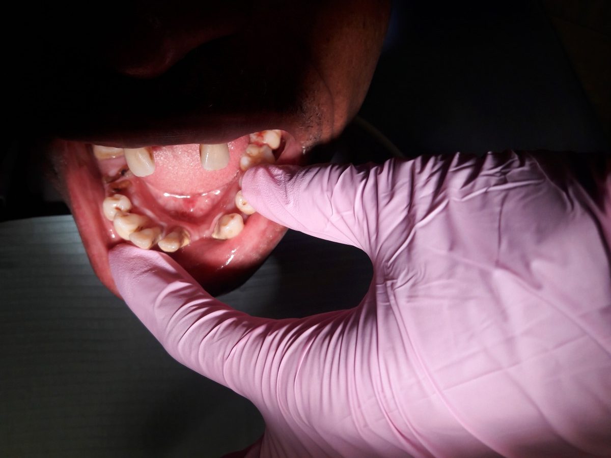
60 year old African male. ASA I. Patient presented with Perio Type IV, caries active, and heavy calculus deposits. **Images of oral cavity were only taken pre-treatment**. Images present above and a link to a comprehensive journal on this patients case below.
Case Study # 3– V.A


74 year old African American woman. ASA II. Vital signs taken: Blood Pressure- 168/75, Pulse- 71. Patient is taken several medications for type 2 Diabetes (last AIC taken 2017, results- 5.7), pulmonary hypertension, anemia, and rheumatoid arthritis. Amlodipine- Enazepril- 10/20 mg, Aspirin 81 mg/q.d., Furosemide 40 mg/q.d., Hydralazine 100 mg, Metformin 1,000 mg/ bid, Metoprolol 200 mg/q.d., Simvastin 40 mg, Tadalafil 20 mg. Patient presented with a cluster of blisters on the attached gingiva of #19-21L, no discomfrot or notice of lesions. Later on in the appointment the lesions were diagnosed as Herpetic lesions. Patient was told to not share anything with anyone that could cause her to transfer the infection.
Case Study # 4– J.F
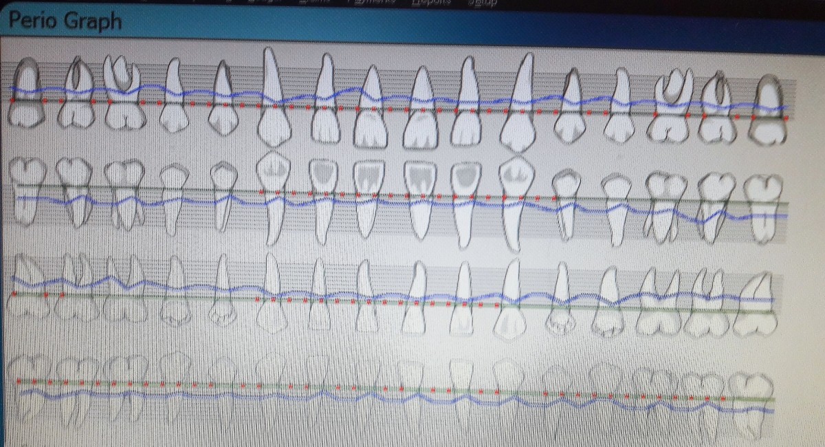
Red dots indicate bleeding points.


24 year old Filipino male, ASA I. Vital signs: Blood Pressure- 140/80, Pulse- 70. Patient presented with severe Erythamatous tissue, rolled gingiva #19 & 31, Bulbous tissue between #7-8 (measured at 3mm by 3mm). Severe Bleeding upon probing, severe bleeding upon exploring, and moderate inflammation. Patient complained of bleeding while flossing. The tissue of his anterior maxillary and mandibular teeth appeared “cherry” red near the gingival margins, and dry. After a thorough inspection of the oral cavity the patient was asked if he is a mouth breather, to which he responded yes. Patient was made aware of tissue condition due to breathing habits. Patient advised to use products with xylitol, increase frequency of flossing, and drink more water to curb the effects of mouth breathing. Images above show patient at revisit appointment, after complete scaling of UR, and LR.

