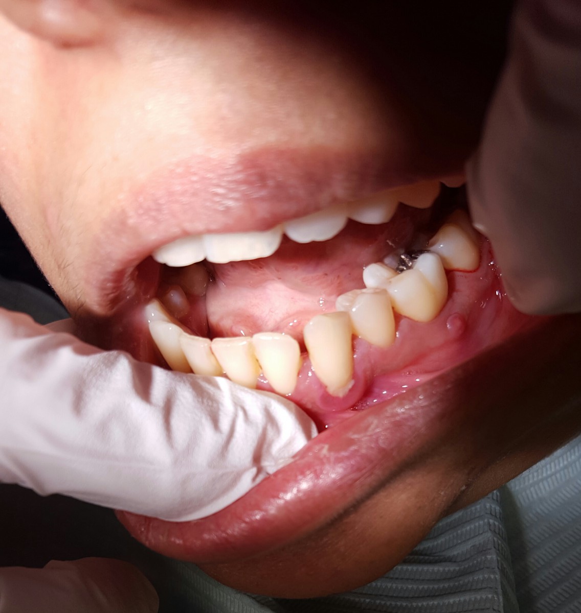
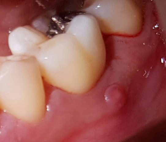
Fistula
Female 37 years, ASA I. BP 96/76 P 83. EO was WNL. IO fistula was found on the attach gingiva of tooth #19. Patient last cleaning was 2015 where she had a filling done on tooth # 19. As IO was conducted the fistula was found and was established to be associated with the restoration on tooth #19, which was broken. Patient presented with red generalized blunted retractable papilla, stippled and non-resilient. Patient had several restoration and missing teeth. Patient was classified as perio type I, with moderate bleeding and inflammation. Patient was disclosed plaque score was 1.2. OHI tooth brushing was taught. Calculus detection was completed and patient was classified a heavy with light stains. Treatment plan was established and executed. First visit all assessment were conducted and tooth #’s 32, 31, and 30 were scaled to completion. Patient was shown the fistula and broken restoration and was advised it needed a dentist to take a look at it. On the 2nd visit all 4 quadrants were scaled to completion, except for tooth #19. # 19 was scaled my the supervising professor with a hand instrument. Patient was given a referral to her dentist for the broken filling and fistula.

Case Study II
Caries
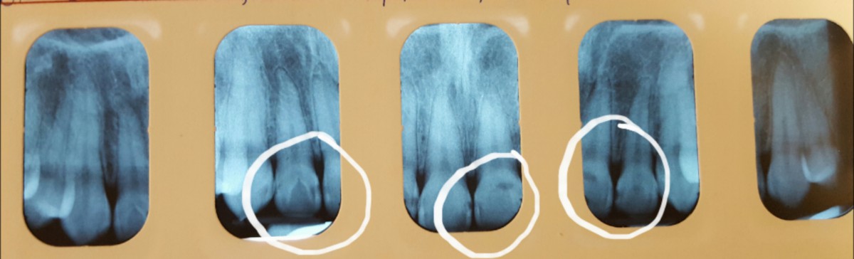
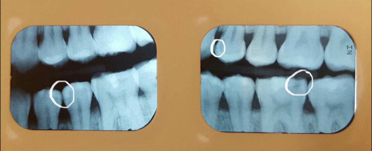
Male 22 years old, ASA I. BP100/66 P 66. EO/IO WNL. Patient presented with chronic inflamed red bulbous gingiva, not resilient or stippled. Patient last cleaning was 7 years ago. Dental charting was completed , patient presented with several caries on posterior and anterior teeth. Patient also had demineralization on all teeth and generalized attrition on anterior teeth. Perio charting was completed and patient was classified as perio type III, with moderate bleeding and severe inflammation. Pt was classified as a heavy. Patient was disclosed and plaque score 2.37, and OHI tooth brushing was taught. On the first visit all the assessment were completed and URQ was scaled. On the second visit FMS was exposed and reviewed with patient. Patient was given a referral to the DDS for caries. The LRQ was scaled that that day and OHI flossing was taught. On the third visit ULQ and LLQ was scaled to completion, OHI tooth brushing was reviewed. PT teeth were polished and fluoride varnish applied.
The radiographs shows the many carious lesions present in the patient’s mouth.